You have no items in your shopping cart.
Cart summary
Item 1 of 2
Item 1 of 2
Anti-FIH-1 Antibody
Catalog Number: orb1473654

| Catalog Number | orb1473654 |
|---|---|
| Category | Antibodies |
| Description | Mouse monoclonal antibody to FIH-1 |
| Species/Host | Mouse |
| Clonality | Monoclonal |
| Tested applications | IF, WB |
| Reactivity | Human |
| Immunogen | KLH-conjugated synthetic peptide encompassing a sequence within the center region of human FIH-1. The exact sequence is proprietary. |
| Antibody Type | Primary Antibody |
| Dilution range | WB (1/1000 - 1/2000), IF/IC (1/10 - 1/50) |
| Form/Appearance | Mouse IgG1 kappa. Liquid in PBS, pH 7.3, 30% glycerol, and 0.01% sodium azide. |
| Conjugation | Unconjugated |
| Target | HIF1AN |
| Entrez | 55662 |
| UniProt ID | Q9NWT6 |
| Source | Mouse |
| Storage | Maintain refrigerated at 2-8°C for up to 2 weeks. For long term storage store at -20°C in small aliquots to prevent freeze-thaw cycles. |
| Buffer/Preservatives | Mouse IgG1 kappa. Liquid in PBS, pH 7.3, 30% glycerol, and 0.01% sodium azide. |
| Alternative names | FIH1; Hypoxia-inducible factor 1-alpha inhibitor; Read more... |
| Note | For research use only |
| Expiration Date | 12 months from date of receipt. |
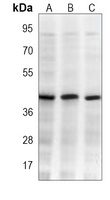
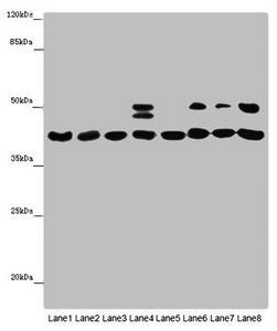
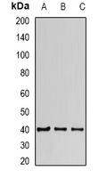
Western blot analysis of FIH-1 expression in 293 (A), Jurkat (B), ZR751 (C) whole cell lysates. (Predicted band size: 40 kD; Observed band size: 40 kD)
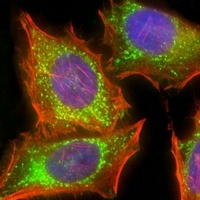
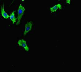
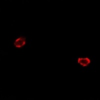
Immunofluorescent analysis of FIH-1 staining in Hela cells. Formalin-fixed cells were permeabilized with 0.1% Triton X-100 in TBS for 5-10 minutes and blocked with 3% BSA-PBS for 30 minutes at room temperature. Cells were probed with the primary antibody in 3% BSA-PBS and incubated overnight at 4 °C in a humidified chamber. Cells were washed with PBST and incubated with a AF488-conjugated secondary antibody (green) in PBS at room temperature in the dark. Phalloidin - AF555 was used to stain Actin filaments (red). DAPI was used to stain the cell nuclei (blue).
- Item 1 of 5
HIF1AN Antibody [orb355183]
ELISA, IF, IHC, IP, WB
Human, Mouse
Rabbit
Polyclonal
Unconjugated
100 μg, 50 μg - Item 1 of 2
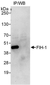
Anti-FIH-1 Antibody [orb340921]
IF, IP, WB
Human, Mouse, Rat
Rabbit
Polyclonal
Unconjugated
200 μl, 100 μl, 50 μl - Item 1 of 2
- Item 1 of 2