You have no items in your shopping cart.
Description
Research Area
Immunology & Inflammation
Images & Validation
−Item 1 of 3
| Tested Applications | ELISA, FC |
|---|---|
| Reactivity | Human |
Key Properties
−| Host | Mouse |
|---|---|
| Clonality | Monoclonal |
| Isotype | IgG2a |
| Clone No. | KS1/4 |
| Immunogen | The original antibody was generated by immunizing BALB/c mice with human adenocarcinoma of the lung cell line UCLA P3. |
| Target | EpCAM |
| Purity | Purified |
| Conjugation | Unconjugated |
Storage & Handling
−| Storage | Maintain refrigerated at 2-8°C for up to 2 weeks. For long term storage store at -20°C in small aliquots to prevent freeze-thaw cycles. |
|---|---|
| Buffer/Preservatives | PBS with 0.02% Proclin 300. |
| Concentration | 1 mg/ml |
| Expiration Date | 12 months from date of receipt. |
| Disclaimer | For research use only |
Alternative Names
−CD326; Ep-CAM; KS1/4 antigen; KSA; Epithelial cell adhesion molecule; Adenocarcinoma-associated antigen; Cell surface glycoprotein Trop-1; Epithelial cell surface antigen; Epithelial glycoprotein; EGP; Epithelial glycoprotein 314; 17-1A-antigen; EGP314; hEGP314; Major gastrointestinal tumor-associated protein GA733-2; Tumor-associated calcium signal transducer 1.
Similar Products
−- Item 1 of 5
- Item 1 of 7
EPCAM Antibody [orb389162]
FC, IF, IHC, WB
Human
Mouse
Monoclonal
Unconjugated
20 μg, 100 μg, 100 μg (without BSA and Azide) - Item 1 of 4
EPCAM Antibody [orb389147]
IHC
Canine, Feline, Human
Mouse
Monoclonal
Unconjugated
20 μg, 100 μg, 100 μg (without BSA and Azide) - Item 1 of 3
- Item 1 of 3
EPCAM Antibody [orb389158]
IHC
Human
Mouse
Monoclonal
Unconjugated
20 μg, 100 μg, 100 μg (without BSA and Azide)
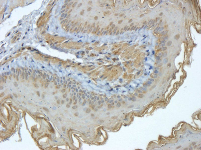
![Anti-EpCAM [KS1/4]](/images/pub/media/catalog/product/NewWebsite/35/orb1089960_1.png)
![Anti-EpCAM [KS1/4]](/images/pub/media/catalog/product/NewWebsite/35/orb1089960_2.png)
![Anti-EpCAM [KS1/4]](/images/pub/media/catalog/product/NewWebsite/35/orb1089960_3.png)


Quality Guarantee
Explore bioreagents carefree to elevate your research. All our products are rigorously tested for performance. If a product does not perform as described on its datasheet, our scientific support team will provide expert troubleshooting, a prompt replacement, or a refund. For full details, please see our Terms & Conditions and Buying Guide. Contact us at [email protected].
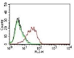
![Anti-EpCAM [KS1/4]](/images/pub/media/catalog/product/NewWebsite/35/orb1089961_1.png)
Flow cytometry using the Anti-EpCAM antibody KS1/4. Paraformaldehyde fixed Caco-2 cells were stained with anti-unknown specificity antibody (orb256458; isotype control, black line) or the rabbit IgG version of KS1/4 (orb1089960, blue line) at a dilution of 1:100 for 1h at RT. After washing, the bound antibody was detected using a goat anti-rabbit IgG AlexaFluor® 488 antibody at a dilution of 1:1000 and cells analyzed using a FACSCanto flow-cytometer.
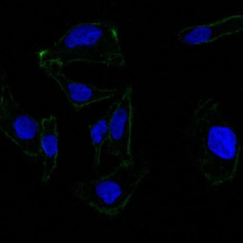
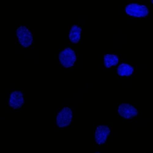
![Anti-EpCAM [KS1/4]](/images/pub/media/catalog/product/NewWebsite/35/orb1089961_2.png)
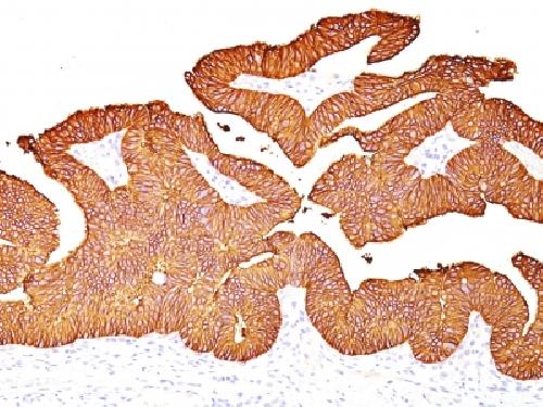
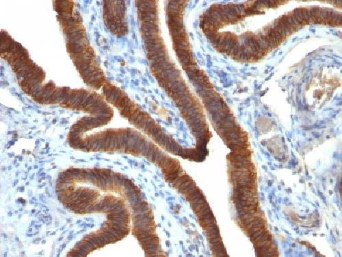
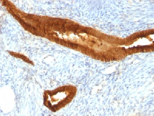
Immunofluorescence staining of Caco-2 cells with anti_EpCAM KS1/4. Immunofluorescence analysis of paraformaldehyde fixed Caco-2 cells stained with the chimeric rabbit IgG version of KS1/4 (orb1089960) (1:100 dilution) for 1h followed by Alexa Fluor® 488 secondary antibody (1:1000 dilution), showing membrane staining. The nuclear stain is DAPI (blue). Panels show from left-right, top-bottom orb1089960, DAPI, merged channels and an isotype control. The isotype control was an unknown specificity antibody (orb256458) followed by staining with Alexa Fluor® 488 secondary antibody.
![Anti-EpCAM [KS1/4]](/images/pub/media/catalog/product/NewWebsite/35/orb1089961_3.png)
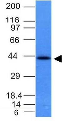
Western Blot using Anti-EpCAM antibody KS1/4. A431(A), Caco-2(B), MCF7(C) and HepG2(D) cell lysates (35 µg protein in RIPA buffer) were resolved on a SDS PAGE gel and blots were probed with the chimeric rabbit version of KS1/4 (orb1089960) at 2 µg/ml before detection using an anti-rabbit secondary antibody. A primary incubation of 1h was used and protein was detected by chemiluminescence.
Quick Database Links
Gene Symbol
EpCAM
UniProt
UniProt Details
− No UniProt data available
Documents Download
Datasheet
Product Information
Request a Document
Protocol Information
FC
Flow Cytometry
ELISA
Enzyme-linked Immunosorbent Assay (EIA)
Anti-EpCAM [KS1/4] (orb1089961)

- 0.0
Based on 0 reviews
Participating in our Biorbyt product reviews program enables you to support fellow scientists by sharing your firsthand experience with our products.
Login to Submit a ReviewAvailable Sizes
Select a size below
Free Secondary Antibody (20 ul)0/0
Please add an antibody product to your cart first.