You have no items in your shopping cart.
Cart summary
Item 1 of 5
Item 1 of 5
AKT1 Antibody (N-term)
Catalog Number: orb1929533

| Catalog Number | orb1929533 |
|---|---|
| Category | Antibodies |
| Description | Purified Rabbit Polyclonal Antibody (Pab) |
| Species/Host | Rabbit |
| Clonality | Polyclonal |
| Clone Number | RB11644 |
| Tested applications | FC, IF, IHC-P, WB |
| Predicted Reactivity | Mouse |
| Reactivity | Human |
| Isotype | Rabbit IgG |
| Dilution range | IF: 1:10~50, WB: 1:1000, WB: 1:1000, IHC-P: 1:50~100, FC: 1:10~50 |
| Form/Appearance | Purified polyclonal antibody supplied in PBS with 0.09% (W/V) sodium azide. This antibody is purified through a protein A column, followed by peptide affinity purification. |
| Conjugation | Unconjugated |
| MW | 55686 Da |
| Target | This AKT1 antibody is generated from rabbits immunized with a KLH conjugated synthetic peptide between 115-144 amino acids from the N-terminal region of human AKT1. |
| UniProt ID | P31749 |
| NCBI | NP_005154.2, NP_001014432.1, NP_001014431.1 |
| Storage | Maintain refrigerated at 2-8°C for up to 2 weeks. For long term storage store at -20°C in small aliquots to prevent freeze-thaw cycles |
| Alternative names | RAC-alpha serine/threonine-protein kinase, Protein Read more... |
| Note | For research use only |
| Expiration Date | 12 months from date of receipt. |
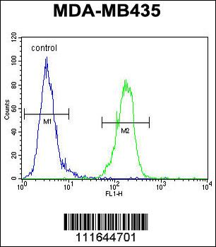
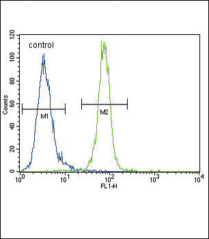
AKT1 Antibody (N-term) flow cytometric analysis of MDA-MB435 cells (right histogram) compared to a negative control cell (left histogram). FITC-conjugated goat-anti-rabbit secondary antibodies were used for the analysis.
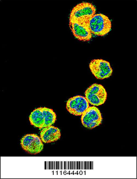
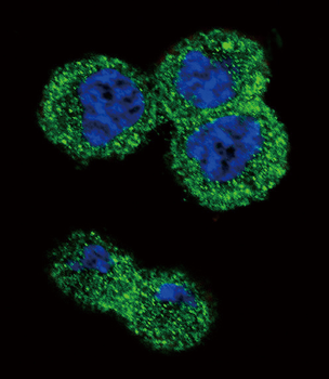
Confocal immunofluorescent analysis of AKT1 Antibody (N-term) with MDA-MB435 cell followed by Alexa Fluor 488-conjugated goat anti-rabbit lgG (green).Actin filaments have been labeled with Alexa Fluor 555 phalloidin (red). DAPI was used to stain the cell nuclear (blue).
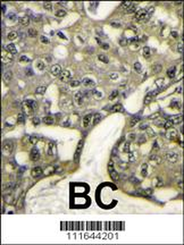
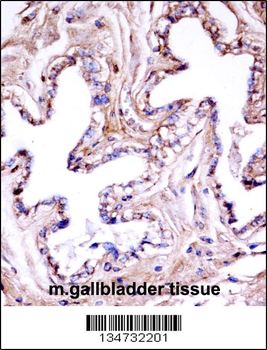
Formalin-fixed and paraffin-embedded human breast carcinoma reacted with AKT1 antibody (N-term), which was peroxidase-conjugated to the secondary antibody, followed by DAB staining. This data demonstrates the use of this antibody for immunohistochemistry; clinical relevance has not been evaluated.
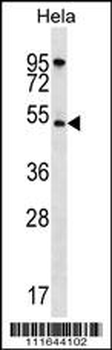
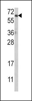
AKT1 Antibody (N-term) western blot analysis in Hela cell line lysates (35 ug/lane). This demonstrates the AKT1 antibody detected the AKT1 protein (arrow).
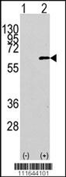
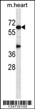
Western blot analysis of AKT1 Antibody (N-term) polyclonal antibody (arrow). 293 cell lysates (2 ug/lane) either nontransfected (Lane 1) or transiently transfected with the AKT1 gene (Lane 2).
- Item 1 of 3
AKT1 Antibody (N-term) [orb1929532]
FC, IF, WB
Mouse, Rat
Human
Rabbit
Polyclonal
Unconjugated
100 μl, 50 μl - Item 1 of 2
Mouse Akt1 Antibody (N-term) [orb1935566]
IHC-P, WB
Human
Mouse
Rabbit
Polyclonal
Unconjugated
100 μl, 50 μl