You have no items in your shopping cart.
Cart summary
Item 1 of 4
Item 1 of 4
UBCH10 Antibody
Catalog Number: orb1246818

| Catalog Number | orb1246818 |
|---|---|
| Category | Antibodies |
| Description | UBCH10 Antibody |
| Target | UBE2C |
| Clonality | Polyclonal |
| Conjugation | Unconjugated |
| Reactivity | Human |
| Form/Appearance | Liquid |
| Concentration | 500 ug/mL |
| Buffer/Preservatives | Supplied at 0.5 mg/ml in Tris saline, 0.02% sodium azide, pH 7.3 with 0.5% bovine serum albumin. Aliquot and store at -20°C. Minimize freezing and thawing. |
| Purification | Purified from goat serum by ammonium sulphate precipitation followed by antigen affinity chromatography using the immunizing peptide. |
| Immunogen | The immunogen for this antibody is: C-QETYSKQVTSQEP |
| UniProt ID | O00762 |
| Tested applications | ELISA, FC, IF, IP, WB |
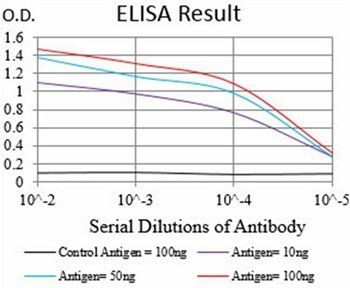
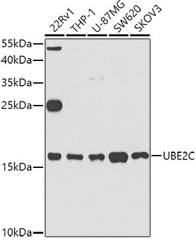
| Application notes | Peptide ELISA: antibody detection limit dilution 1:32000.Western Blot:Approx.19KDa band observed in lysates of cell lines HeLa and HEK293 (calculated MW of 19.7kDa according to NP_008950.1). Recommended concentration: 1-3ug/ml. Primary incubation 1 hour at room temperature.Immunofluorescence: Strong expression of the protein seen in the cytoplasm of U2OS and MCF7 cells. Recommended concentration: 10ug/ml. Flow Cytometry: Flow cytometric analysis of HeLa cells. Recommended concentration: 10ug/ml. Immunoprecipitation: 20KDa band precipitated from mitotic HeLa whole cell lysates using protein-G dynabeads. |
| Antibody Type | Primary Antibody |
| Storage | Maintain refrigerated at 2-8°C for up to 2 weeks. For long term storage store at -20°C in small aliquots to prevent freeze-thaw cycles. |
| Alternative names | UBE2C, UBCH10, dJ447F3.2, ubiquitin-conjugating en Read more... |
| Note | For research use only |
| NCBI | NP_861518.1, NP_861515.1, NP_001268670.1, NP_008950.1, NP_861516.1, NP_861517.1 |

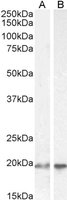
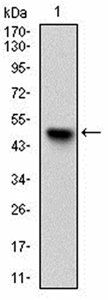
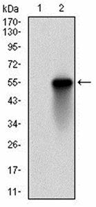
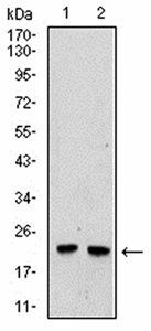
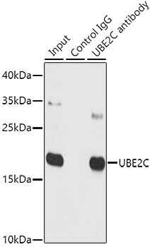
orb1246818 (2 ug/ml) staining of HEK293 (A) and HeLa (B) cell lysate (35 ug protein in RIPA buffer). Detected by chemiluminescence.
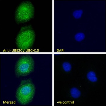
orb1246818 Immunofluorescence analysis of paraformaldehyde fixed U2OS cells, permeabilized with 0.15% Triton. Primary incubation 1hr (10 ug/ml) followed by Alexa Fluor 488 secondary antibody (4 ug/ml), showing cytoplasmic /Plasma Membrane staining.
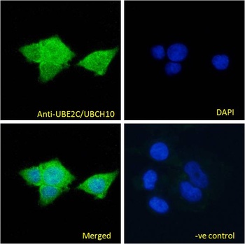
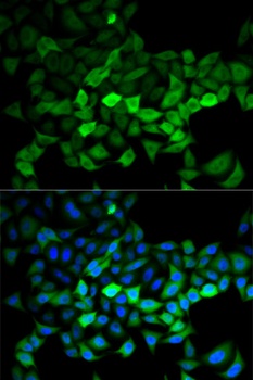
orb1246818 Immunofluorescence analysis of paraformaldehyde fixed MCF7 cells, permeabilized with 0.15% Triton. Primary incubation 1hr (10 ug/ml) followed by Alexa Fluor 488 secondary antibody (4 ug/ml), showing cytoplasmic staining. The nuclear stain is DAPI.
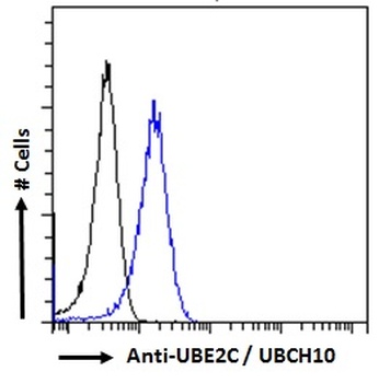
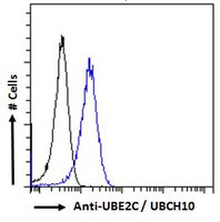
orb1246818 Flow cytometric analysis of paraformaldehyde fixed HeLa cells (blue line), permeabilized with 0.5% Triton. Primary incubation 1hr (10 ug/ml) followed by Alexa Fluor 488 secondary antibody (2 ug/ml). IgG control: Unimmunized goat IgG (black line).
- Item 1 of 4
Goat anti-UBE2C / UBCH10 Antibody [orb18433]
ELISA, FC, IF, IP, WB
Human
Goat
Polyclonal
Unconjugated
100 μg - Item 1 of 6
- Item 1 of 5
- Item 1 of 3
- Item 1 of 3


![Anti-UBE2C [SAIC-41A-1]](/images//pub/media/catalog/product/NewWebsite/35/orb1089976_1.png)
![Anti-UBE2C [SAIC-41A-1]](/images/pub/media/catalog/product/NewWebsite/35/orb1089976_2.png)
![Anti-UBE2C [SAIC-41A-1]](/images/pub/media/catalog/product/NewWebsite/35/orb1089976_3.png)