You have no items in your shopping cart.
Rabbit anti-MafA Recombinant Monoclonal Antibody
SKU: orb1519834
Description
Images & Validation
−Item 1 of 7
| Tested Applications | ICC, IHC, WB |
|---|---|
| Dilution range | WB - 1:1000; IHC - 1:100 to 1:500. Epitope retrieval with citrate buffer pH6.0 is recommended for FFPE tissue sections. Zinc-fixative (JB fix) is recommended to enhance staining.; ICC - 1:100 to 1:500. Epitope retrieval with citrate buffer pH6.0 is recommended for FFPE cell sections.; IHC-IF - 1:100 to 1:500. Epitope retrieval with citrate buffer pH6.0 is recommended for FFPE tissue sections. Zinc-fixative (JB fix) is recommended to enhance staining. |
| Reactivity | Human, Mouse |
| Application Notes |
Key Properties
−| Antibody Type | Primary Antibody |
|---|---|
| Host | Rabbit |
| Clonality | Recombinant |
| Clone No. | BLR067G |
| Immunogen | Between 125 and 175 |
| Target | MafA |
Storage & Handling
−| Storage | 2 - 8°C |
|---|---|
| Form/Appearance | Whole IgG |
| Buffer/Preservatives | Borate Buffered Saline (BBS) pH 8.2 with 0.09% Sodium Azide, rAlbumin-Free |
| Concentration | 1000 µg/ml |
| Disclaimer | For research use only |
Alternative Names
−v-maf avian musculoaponeurotic fibrosarcoma oncogene homolog A; V-maf musculoaponeurotic fibrosarcoma oncogene homolog A; transcription factor RIPE3b1; transcription factor MafA; RIPE3b1 factor; RIPE3b1; pancreatic beta-cell-specific transcriptional activator; INSDM; hMafA
Similar Products
−- Item 1 of 7
Rabbit anti-MafA Recombinant Monoclonal Antibody [orb1519835]
ICC, IHC, WB
Human, Mouse
Rabbit
Recombinant
Unconjugated
100 μl


Quality Guarantee
Explore bioreagents carefree to elevate your research. All our products are rigorously tested for performance. If a product does not perform as described on its datasheet, our scientific support team will provide expert troubleshooting, a prompt replacement, or a refund. For full details, please see our Terms & Conditions and Buying Guide. Contact us at [email protected].

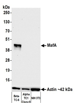
Detection of mouse MafA by western blot. Samples: Whole cell lysate (10 µg) from Beta-TC-6, AlphaTC1 Clone 9, and NIH 3T3 cells prepared using NETN lysis buffer. Antibody: Rabbit anti-MafA recombinant monoclonal antibody (orb1519834) used at 1:1000. Secondary: HRP-conjugated goat anti-rabbit IgG. Detection: Chemiluminescence with an exposure time of 10 seconds. Lower Panel: Rabbit anti-Actin recombinant monoclonal antibody.

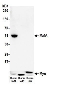
Detection of human MafA by western blot. Samples: Whole cell lysate (10 µg) from HEK293T transfected with myc tagged Human MafA, Human MafB, and Human cMaf prepared using NETN lysis buffer. Antibody: Rabbit anti-MafA recombinant monoclonal antibody (orb1519834) used at 1:1000. Secondary: HRP-conjugated goat anti-rabbit IgG. Detection: Chemiluminescence with an exposure time of 30 seconds. Lower Panel: Rabbit recombinant monoclonal antibody to Myc-tag.

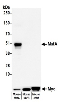
Detection of mouse MafA by western blot. Samples: Whole cell lysate (10 µg) from HEK293T transfected with myc tagged mouse MafA, Human MafB, and Human cMaf prepared using NETN lysis buffer. Antibody: Rabbit anti-MafA recombinant monoclonal antibody (orb1519834) used at 1:1000. Secondary: HRP-conjugated goat anti-rabbit IgG. Detection: Chemiluminescence with an exposure time of 30 seconds. Lower Panel: Rabbit recombinant monoclonal antibody to Myc-tag.
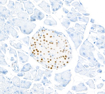
Detection of mouse MafA by immunohistochemistry. Sample: FFPE section of mouse pancreas (JB fixation). Antibody: Rabbit anti-MafA recombinant monoclonal antibody (orb1519834). Secondary: HRP-conjugated goat anti-rabbit IgG.
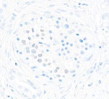
Detection of human MafA by immunohistochemistry. Sample: FFPE section of human pancreatic tumor. Antibody: Rabbit anti-MafA recombinant monoclonal antibody (orb1519834). Secondary: HRP-conjugated goat anti-rabbit IgG.

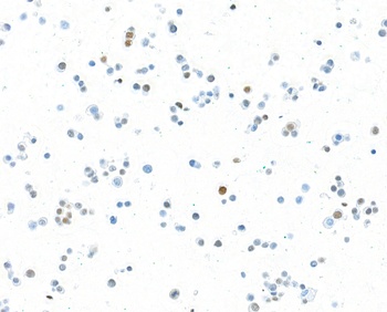
Detection of mouse MafA by immunohistochemistry. Sample: FFPE section of mouseBeta-TC-6 cells. Antibody: Rabbit anti-MafA recombinant monoclonal antibody (orb1519834). Secondary: HRP-conjugated goat anti-rabbit IgG.
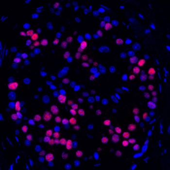
Detection of human MafA (red) by immunohistochemistry. Sample: FFPE section of human pancreatic carcinoma. Antibody: Rabbit anti-MafA recombinant monoclonal antibody (orb1519834) used at 1:250. Secondary: HRP-conjugated goat anti-rabbit IgG. Substrate: Opal™. Counterstain: DAPI (blue).
Quick Database Links
UniProt Details
− No UniProt data available
NCBI Reference Sequences
−Associated Accession Numbers
Curated reference sequences for the gene transcript and protein product| Protein | NP_963883.2 |
|---|
Documents Download
Datasheet
Product Information
Request a Document
Protocol Information
WB
Western Blot (IB, immunoblot)
IHC
Immunohistochemistry
ICC
Immunocytochemistry
Rabbit anti-MafA Recombinant Monoclonal Antibody (orb1519834)

- 0.0
Based on 0 reviews
Participating in our Biorbyt product reviews program enables you to support fellow scientists by sharing your firsthand experience with our products.
Login to Submit a ReviewAvailable Sizes
Select a size below