You have no items in your shopping cart.
PARK7/DJ1 Rabbit Polyclonal Antibody
SKU: orb10543
Featured
Description
Images & Validation
−Item 1 of 7
| Tested Applications | FC, IF, IHC-Fr, IHC-P, WB |
|---|---|
| Dilution range | WB=1:500-2000, IHC-P=1:100-500, IHC-F=1:100-500, IF=1:100-500, Flow-Cyt=0.2μg/Test |
| Reactivity | Human, Mouse, Rat |
| Predicted Reactivity | Bovine, Equine, Porcine |
Related Conjugates & Formulations
−Key Properties
−| Antibody Type | Primary Antibody |
|---|---|
| Host | Rabbit |
| Clonality | Polyclonal |
| Isotype | IgG |
| Immunogen | KLH conjugated synthetic peptide derived from human CAP1 (101-189/189aa) |
| Target | PARK7 |
| Molecular Weight | 20 kDa |
| Purification | Affinity purified by Protein A |
Storage & Handling
−| Storage | Maintain refrigerated at 2-8°C for up to 2 weeks. For long term storage store at -20°C in small aliquots to prevent freeze-thaw cycles. |
|---|---|
| Form/Appearance | Liquid |
| Buffer/Preservatives | 0.01M TBS (pH7.4) with 1% rAlbumin, 0.02% Proclin300 and 50% Glycerol. |
| Concentration | 1mg/ml |
| Disclaimer | For research use only |
Alternative Names
−PARK7; PARK-7; Parkinson disease protein 7; Park7 protein; CAP1; DJ-1; DJ 1; SP22; Protein DJ-1; Oncogene DJ1; FLJ27376; Park 7; Parkinson disease(autosomal recessive early onset) 7; RNA binding protein regulatory subunit; RS; SP22; CAP1_HUMAN.
Similar Products
−
PARK7/DJ1 Rabbit Polyclonal Antibody (HRP) [orb479524]
IHC-Fr, IHC-P, WB
Bovine, Equine, Porcine
Human, Mouse, Rat
Rabbit
Polyclonal
HRP
100 μlPARK7/DJ1 Rabbit Polyclonal Antibody (Cy7) [orb1052436]
FC, IF
Bovine, Equine, Porcine
Human, Mouse, Rat
Rabbit
Polyclonal
Cy7
100 μlPARK7/DJ1 Rabbit Polyclonal Antibody (Cy5.5) [orb1052408]
FC, IF
Bovine, Equine, Porcine
Human, Mouse, Rat
Rabbit
Polyclonal
Cy5.5
100 μlPARK7/DJ1 Rabbit Polyclonal Antibody (RBITC) [orb1052649]
FC, IF
Bovine, Equine, Porcine
Human, Mouse, Rat
Rabbit
Polyclonal
RBITC
100 μlPARK7/DJ1 Rabbit Polyclonal Antibody (Cy3) [orb987826]
FC, IF
Bovine, Equine, Porcine
Human, Mouse, Rat
Rabbit
Polyclonal
Cy3
100 μl
Quality Guarantee
Explore bioreagents carefree to elevate your research. All our products are rigorously tested for performance. If a product does not perform as described on its datasheet, our scientific support team will provide expert troubleshooting, a prompt replacement, or a refund. For full details, please see our Terms & Conditions and Buying Guide. Contact us at [email protected].
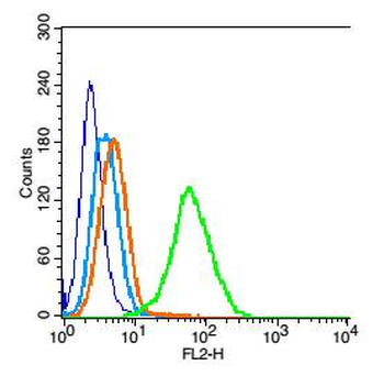
Blank control: 293T cells (blue). Primary Antibody: Rabbit Anti-PARK7/CAP1 antibody (orb10543), Dilution: 0.2 µg in 100 µl 1X PBS containing 0.5% BSA, Isotype Control Antibody: Rabbit IgG (orange), used under the same conditions. Secondary Antibody: Goat anti-rabbit IgG-PE (white blue), Dilution: 1:200 in 1 X PBS containing 0.5% BSA. Protocol, Primary antibody (orb10543, 0.2 µg/1x10^6 cells) were incubated for 30 min on the ice, followed by 1 X PBS containing 0.5% BSA + 1 0% goat serum (15 min) to block non-specific protein-protein interactions. Then the Goat Anti-rabbit IgG/PE antibody was added into the blocking buffer mentioned above to react with the primary antibody at 1/200 dilution for 30 min on ice. Acquisition of 20000 events was performed.
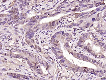
Paraformaldehyde-fixed, paraffin embedded (human colon carcinoma), Antigen retrieval by boiling in sodium citrate buffer (pH6.0) for 15 min, Block endogenous peroxidase by 3% hydrogen peroxide for 20 minutes, Blocking buffer (normal goat serum) at 37°C for 30 min, Antibody incubation with (PARK7) Polyclonal Antibody, Unconjugated (orb10543) at 1:200 overnight at 4°C, followed by operating according to SP Kit (Rabbit) instructionsand DAB staining.
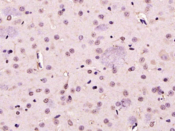
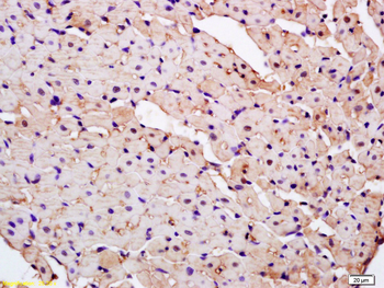
Paraformaldehyde-fixed, paraffin embedded (rat brain), Antigen retrieval by boiling in sodium citrate buffer (pH6.0) for 15 min, Block endogenous peroxidase by 3% hydrogen peroxide for 20 minutes, Blocking buffer (normal goat serum) at 37°C for 30 min, Antibody incubation with (PARK7) Polyclonal Antibody, Unconjugated (orb10543) at 1:200 overnight at 4°C, followed by operating according to SP Kit (Rabbit) instructionsand DAB staining.
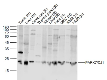
Sample: Lane 1: Testis (Mouse) Lysate at 40 ug, Lane 2: Liver (Mouse) Lysate at 40 ug, Lane 3: Cerebrum (Rat) Lysate at 40 ug, Lane 4: Thyroid gland Rat) Lysate at 40 ug, Lane 5: Kidney (Rat) Lysate at 40 ug, Lane 6: Liver (Rat) Lysate at 40 ug, Lane 7: Hela (Human) Cell Lysate at 30 ug, Lane 8: U937 (Human) Cell Lysate at 30 ug, Lane 9: K562 (Human) Cell Lysate at 30 ug, Lane 10: HL60 (Human) Cell Lysate at 30 ug, Primary: Anti-PARK7/DJ1 (orb10543) at 1/1000 dilution, Secondary: IRDye800CW Goat Anti-Rabbit IgG at 1/20000 dilution, Predicted band size: 22 kD, Observed band size: 22 kD.
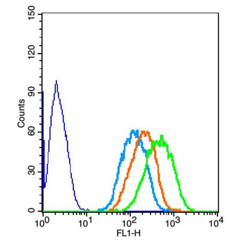
The blue histogram is unstained cells (Hepg2 cells), concentration 1:50, The Wathet Blue histogram is cells stained with secondary antibody alone. The Orange histogram is cells stained with rabbit IgG isotype control antibody, plus secondary antibody. The green histogram is cells stained with Rabbit Anti-PARK7/CAP1 antibody (orb10543) plus secondary antibody.
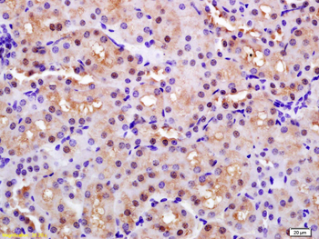
Tissue/Cell: rat kidney tissue, 4% Paraformaldehyde-fixed and paraffin-embedded, Antigen retrieval: citrate buffer (0.01M, pH6.0), Boiling bathing for 15 min, Block endogenous peroxidase by 3% Hydrogen peroxide for 30 min, Blocking buffer (normal goat serum) at 37°C for 20 min, Incubation: Anti-CAP1/PARK7 Polyclonal Antibody, Unconjugated (orb10543) 1:200, overnight at 4°C, followed by conjugation to the secondary antibody and DAB staining.
Tissue/Cell: rat kidney tissue, 4% Paraformaldehyde-fixed and paraffin-embedded, Antigen retrieval: citrate buffer (0.01M, pH6.0), Boiling bathing for 15 min, Block endogenous peroxidase by 3% Hydrogen peroxide for 30 min, Blocking buffer (normal goat serum) at 37°C for 20 min, Incubation: Anti-CAP1/PARK7 Polyclonal Antibody, Unconjugated (orb10543) 1:200, overnight at 4°C, followed by conjugation to the secondary antibody and DAB staining.
Quick Database Links
Gene Symbol
PARK7
UniProt
UniProt Details
− No UniProt data available
Documents Download
Datasheet
Product Information
Request a Document
Protocol Information
WB
Western Blot (IB, immunoblot)
IHC-P
Immunohistochemistry Paraffin
IHC-Fr
Immunohistochemistry Frozen
FC
Flow Cytometry
IF
Immunofluorescence
PARK7/DJ1 Rabbit Polyclonal Antibody (orb10543)

- 0.0
Based on 0 reviews
Participating in our Biorbyt product reviews program enables you to support fellow scientists by sharing your firsthand experience with our products.
Login to Submit a ReviewAvailable Sizes
Select a size below