You have no items in your shopping cart.
Cart summary
Item 1 of 3
Item 1 of 3
p53 Antibody: FITC
Catalog Number: orb377406

| Catalog Number | orb377406 |
|---|---|
| Category | Antibodies |
| Description | Rabbit polyclonal antibody against p53 conjugated to FITC. |
| Species/Host | Rabbit |
| Clonality | Polyclonal |
| Tested applications | ICC, IF, IHC |
| Reactivity | Human |
| Immunogen | Synthetic peptide from the C-terminal of human tumor protein p53. |
| Concentration | 1 mg/ml |
| Dilution range | WB (1:1000); ICC/IF (1:100) |
| Conjugation | FITC |
| MW | 53kDa |
| Target | p53 |
| Entrez | 7157 |
| UniProt ID | P04637 |
| NCBI | NP_000537.3 |
| Storage | Conjugated antibodies should be stored according to the product label |
| Buffer/Preservatives | 640.91mM DMSO, 136.36mM Ethanolamine, and 9.09mM Sodium Bicarbonate in 90.9% PBS |
| Alternative names | TP53 antibody, Tumor Protein 53 antibody, BCC7 ant Read more... |
| Note | For research use only |
| Application notes | A 1:1000 dilution of SPC-682 was sufficient for detection of p53 on HeLa cell lysates using Goat anti-rabbit IgG:HRP as the secondary antibody. |
| Expiration Date | 12 months from date of receipt. |

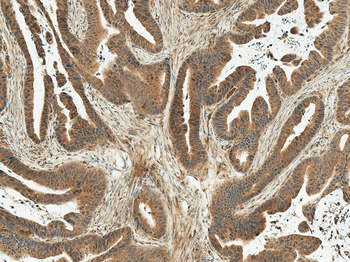
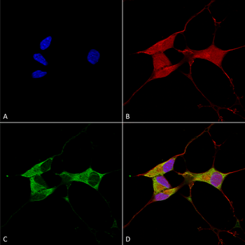
Immunocytochemistry/Immunofluorescence analysis using Rabbit Anti-p53 Polyclonal Antibody. Tissue: Cervical cancer cell line (HeLa). Species: Human. Fixation: 4% Formaldehyde for 15 min at RT. Primary Antibody: Rabbit Anti-p53 Polyclonal Antibody at 1:100 for 60 min at RT. Secondary Antibody: Goat Anti-Rabbit ATTO 488 at 1:100 for 60 min at RT. Counterstain: DAPI (blue) nuclear stain at 1:5000 for 5 min RT. Localization: Cytoplasm, PML body, Endoplasmic Reticulum. Magnification: 40X. (A) DAPI (blue) nuclear stain (B) Phalloidin Texas Red F-Actin stain (C) p53 Antibody (D) Composite.
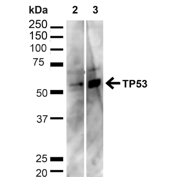
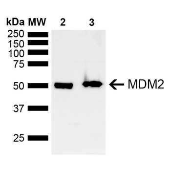
Western blot analysis of Human HeLa and HEK293T cell lysates showing detection of ~43.7kDa p53 protein using Rabbit Anti-p53 Polyclonal Antibody. Lane 1: MW Ladder. Lane 2: Human HeLa (20 μg). Lane 3: Human 293T (20 μg). Load: 20 μg. Block: 5% milk + TBST for 1 hour at RT. Primary Antibody: Rabbit Anti-p53 Polyclonal Antibody at 1:1000 for 1 hour at RT. Secondary Antibody: Goat Anti-Rabbit: HRP at 1:2000 for 1 hour at RT. Color Development: TMB solution for 12 min at RT. Predicted/Observed Size: ~43.7kDa.
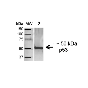
Western blot analysis of Human A431 showing detection of ~43.7kDa p53 protein using Rabbit Anti-p53 Polyclonal Antibody. Lane 1: MW Ladder. Lane 2: Human A431 (20 μg). Load: 20 μg. Block: 5% milk + TBST for 1 hour at RT. Primary Antibody: Rabbit Anti-p53 Polyclonal Antibody at 1:1000 for 1 hour at RT. Secondary Antibody: Goat Anti-Rabbit: HRP at 1:2000 for 1 hour at RT. Color Development: TMB solution for 12 min at RT. Predicted/Observed Size: ~43.7kDa.
- Item 1 of 1
- Item 1 of 2

CDKN2A/p16-INK4a Rabbit Polyclonal Antibody (FITC) [orb16133]
IF
Porcine
Human
Rabbit
Polyclonal
FITC
100 μl- Item 1 of 1