You have no items in your shopping cart.
Cart summary
Item 1 of 4
Item 1 of 4
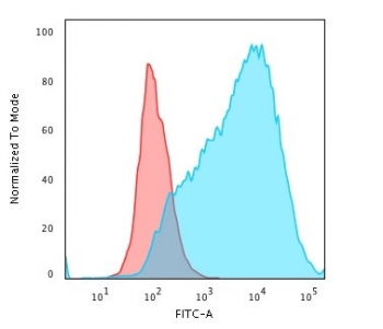
MUC1 Antibody / Mucin-1
Catalog Number: orb2636783

| Catalog Number | orb2636783 |
|---|---|
| Category | Antibodies |
| Description | Mucin-1 may provide a protective layer on epithelial cells against bacterial and enzyme attack. In immunohistochemical assays, it superbly stains routine formalin/paraffin carcinomas. Anti-Mucin-1 antibody is a useful marker for staining many carcinomas. It stains normal and neoplastic cells from various tissues, including mammary epithelium, sweat glands and colorectal carcinoma. Hepatocellular carcinoma, adrenal carcinoma and embryonal carcinomas are consistently Mucin-1 negative, so keratin positivity with negative Mucin-1 favors one of these tumors. Mucin-1 is frequently positive in meningioma, which can be useful when distinguishing it from other intracranial neoplasms such as schwannomas. Antibody to Mucin-1 is useful as a pan-epithelial marker for detecting early metastatic loci of carcinoma in bone marrow or liver. |
| Species/Host | Rabbit |
| Clonality | Recombinant |
| Clone Number | MUC1/4416R |
| Tested applications | IHC-P, WB |
| Reactivity | Human |
| Isotype | Rabbit IgG |
| Immunogen | Recombinant full-length human MUC1 protein was used as the immunogen for the recombinant Mucin-1 antibody. |
| Antibody Type | Primary Antibody |
| Dilution range | Western blot: 1-2ug/ml,Immunohistochemistry (FFPE): 1-2ug/ml |
| Purity | Protein A/G affinity |
| Conjugation | Unconjugated |
| Formula | 0.2 mg/ml in 1X PBS with 0.1 mg/ml BSA (US sourced), 0.05% sodium azide |
| Hazard Information | This recombinant Mucin-1 antibody is available for research use only. |
| UniProt ID | P15941 |
| Storage | Maintain refrigerated at 2-8°C for up to 2 weeks. For long term storage store at -20°C in small aliquots to prevent freeze-thaw cycles. |
| Buffer/Preservatives | 0.2 mg/ml in 1X PBS with 0.1 mg/ml rAlbumin (US sourced), 0.05% sodium azide |
| Note | For research use only |
| Application notes | Optimal dilution of the recombinant Mucin-1 antibody should be determined by the researcher. |
| Expiration Date | 12 months from date of receipt. |
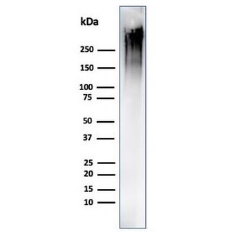
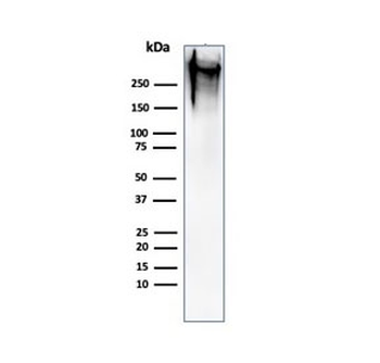
Western blot testing of human MCF-7 cell lysate using recombinant Mucin-1 antibody (clone MUC1/4416R). This glycoprotein is commonly visualized between 120~500 kDa.
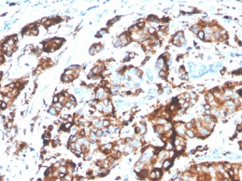
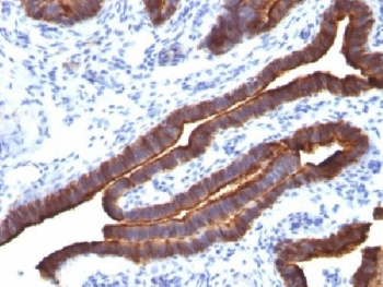
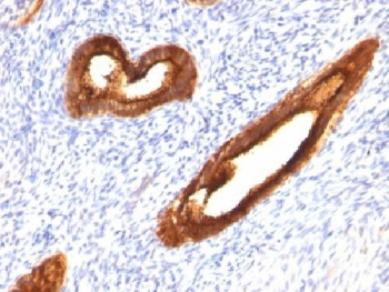
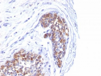
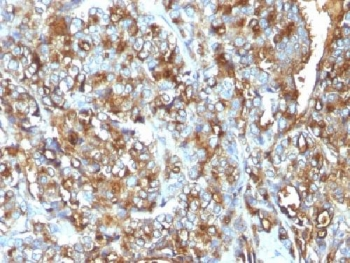
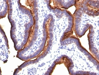
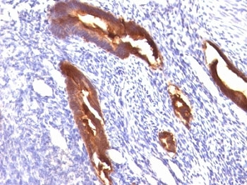
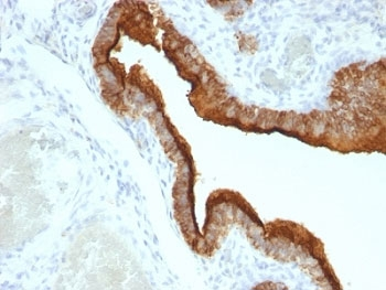
IHC staining of FFPE human breast carcinoma tissue with recombinant Mucin-1 antibody (clone MUC1/4416R). HIER: boil tissue sections in pH9 10mM Tris with 1mM EDTA for 20 min and allow to cool before testing.
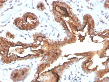
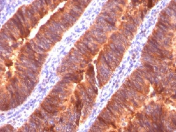
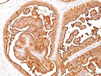
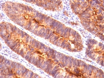
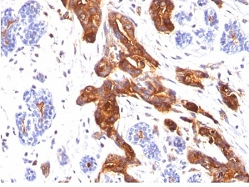
IHC staining of FFPE human breast carcinoma tissue with recombinant Mucin-1 antibody (clone MUC1/4416R). HIER: boil tissue sections in pH9 10mM Tris with 1mM EDTA for 20 min and allow to cool before testing.
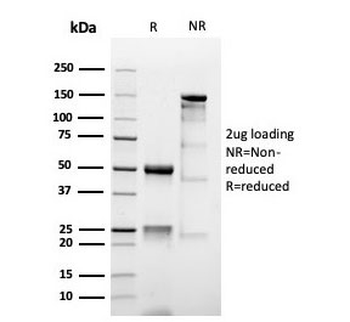
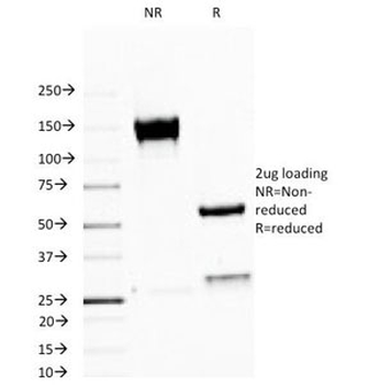
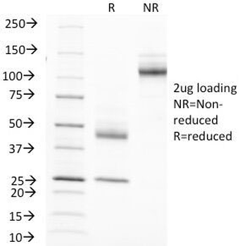
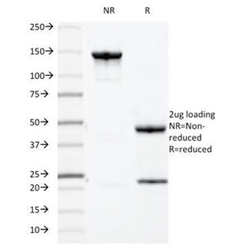
SDS-PAGE analysis of purified, BSA-free recombinant Mucin-1 antibody (clone MUC1/4416R) as confirmation of integrity and purity.
- Item 1 of 8
- Item 1 of 8
- Item 1 of 7
- Item 1 of 7
- Item 1 of 5