You have no items in your shopping cart.
MSH6 Antibody
SKU: orb1261737
Featured
Description
Images & Validation
−Item 1 of 6
| Tested Applications | IF, IHC, WB |
|---|---|
| Reactivity | Human, Monkey, Mouse |
| Application Notes |
Key Properties
−| Antibody Type | Primary Antibody |
|---|---|
| Host | Rabbit |
| Clonality | Polyclonal |
| Isotype | IgG |
| Immunogen | Recombinant fusion protein containing a sequence corresponding to amino acids 1-290 of human MSH6 (NP_000170.1). |
| Target | MSH6 |
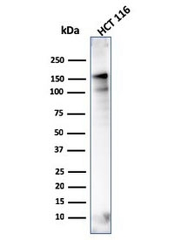
| Molecular Weight | Observed: 160kDa |
| Purification | Affinity purification |
| Conjugation | Unconjugated |
Storage & Handling
−| Storage | Maintain refrigerated at 2-8°C for up to 2 weeks. For long term storage store at -20°C in small aliquots to prevent freeze-thaw cycles. |
|---|---|
| Buffer/Preservatives | Liquid |
| Concentration | batch dependent |
| Disclaimer | For research use only |
Alternative Names
−GTBP, GTMBP, HNPCC5, HSAP, p16DNA mismatch repair protein Msh6, G/T mismatch-binding protein, mutS protein homolog 6, mutS-alpha 160 kDa subunit, sperm-associated protein
Similar Products
−- Item 1 of 7
- Item 1 of 6
MSH6 Recombinant Rabbit Monoclonal Antibody [orb1151975]
FC, IF, IHC-Fr, IHC-P, WB
Mouse, Rat
Human, Mouse, Rat
Rabbit
Recombinant
Unconjugated
25 μl, 100 μl, 50 μl - Item 1 of 7
- Item 1 of 7
- Item 1 of 7


Quality Guarantee
Explore bioreagents carefree to elevate your research. All our products are rigorously tested for performance. If a product does not perform as described on its datasheet, our scientific support team will provide expert troubleshooting, a prompt replacement, or a refund. For full details, please see our Terms & Conditions and Buying Guide. Contact us at [email protected].
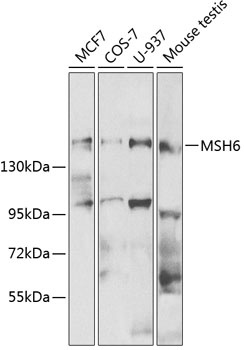
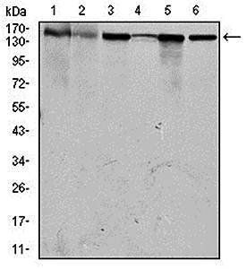
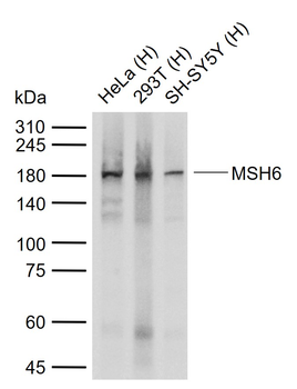
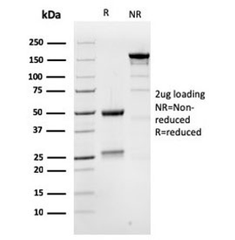
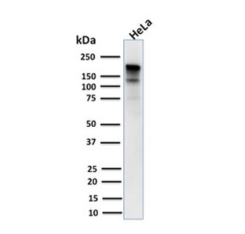
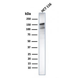
Western blot analysis of extracts of various cell lines, using MSH6 antibody at 1:500 dilution.
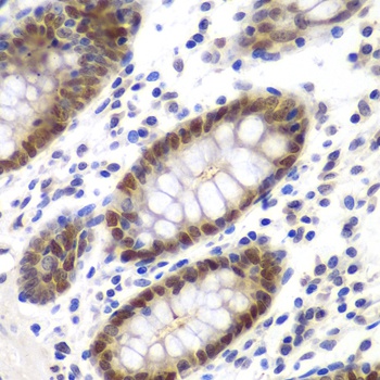
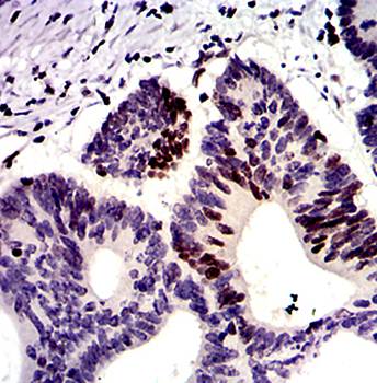
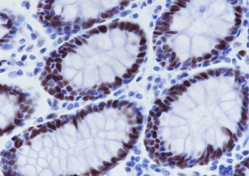
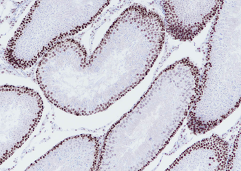
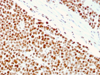
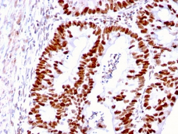
Immunohistochemistry of paraffin-embedded human colon using MSH6 antibody at dilution of 1:100 (40x lens).
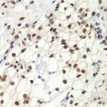
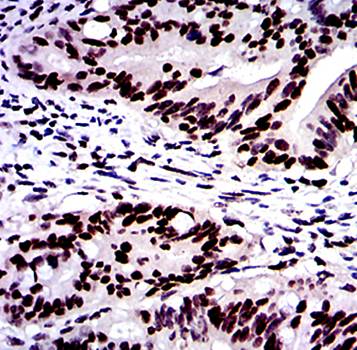
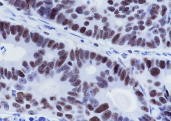
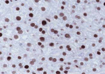
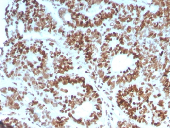
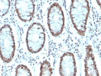
Immunohistochemistry of paraffin-embedded human kidney cancer using MSH6 antibody at dilution of 1:100 (40x lens).
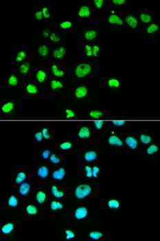
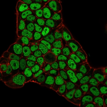
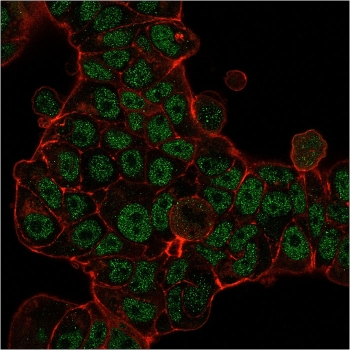
Immunofluorescence analysis of HeLa cells using MSH6 antibody. Blue: DAPI for nuclear staining.
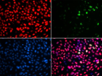
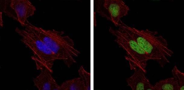
Immunofluorescence analysis of GFP-RNF168 transgenic U2OS cells using MSH6 antibody. Green:GFP-RNF168 fusion protein expression for DNA damage marker. Blue: DAPI for nuclear staining. RNF168 (GFP) can be used to mark cells damaged by UV-A laser for they always gather around DNA damage region.
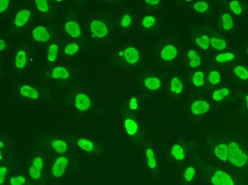
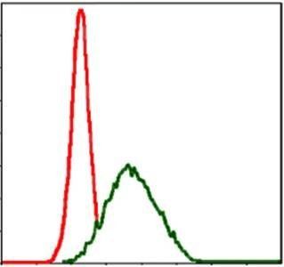
Immunofluorescence analysis of A549 cells using MSH6 antibody.
Quick Database Links
Gene Symbol
MSH6
UniProt
UniProt Details
− No UniProt data available
Documents Download
Datasheet
Product Information
Request a Document
Protocol Information
WB
Western Blot (IB, immunoblot)
IHC
Immunohistochemistry
IF
Immunofluorescence
MSH6 Antibody (orb1261737)

- 0.0
Based on 0 reviews
Participating in our Biorbyt product reviews program enables you to support fellow scientists by sharing your firsthand experience with our products.
Login to Submit a ReviewAvailable Sizes
Select a size below