You have no items in your shopping cart.
Page Not Found
Cart summary
Item 1 of 3
Item 1 of 3
Human IgM mu chain Antibody
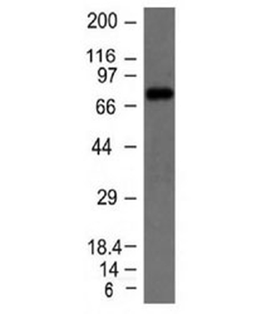
Catalog Number: orb750701

| Catalog Number | orb750701 |
|---|---|
| Category | Antibodies |
| Description | Human IgM mu chain |
| Species/Host | Rabbit |
| Clonality | Polyclonal |
| Tested applications | ELISA, IHC, WB |
| Reactivity | Human |
| Isotype | Antiserum |
| Immunogen | Human IgM mu heavy chain |
| Antibody Type | Secondary Antibody |
| Concentration | 90 mg/mL |
| Dilution range | ELISA: 1:20,000 - 1:100,000, IHC: 1:1,000 - 1:5,000, WB: 1:2,000 - 1:10,000 |
| Form/Appearance | Lyophilized |
| Purity | This product was prepared from monospecific antiserum by a delipidation and defibrination. Assay by immunoelectrophoresis resulted in a single precipitin arc against anti-rabbit serum, Human IgM and Human Serum. No reaction was observed against Human IgG. |
| Conjugation | Unconjugated |
| Storage | Store vial at 4° C prior to restoration. For extended storage aliquot contents and freeze at -20° C or below. Avoid cycles of freezing and thawing. Centrifuge product if not completely clear after standing at room temperature. This product is stable for several weeks at 4° C as an undiluted liquid. Dilute only prior to immediate use. |
| Buffer/Preservatives | 0.01% (w/v) Sodium Azide |
| Alternative names | rabbit anti-Human IgM mu chain Antibody, rabbit an Read more... |
| Note | For research use only |
| Application notes | Anti-Human IgM (mu heavy chain) is designed for immunofluorescence microscopy, fluorescence based plate assays (FLISA) and fluorescent western blotting. This product is also suitable for multiplex analysis, including multicolor imaging, utilizing various commercial platforms. |
| Expiration Date | 12 months from date of receipt. |
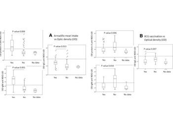
ELISA results using Rabbit Anti-Human IgM. Impact of behavioral variables on M. leprae infection levels. Anti-natural octyl disaccharide-leprosy IDRI diagnostic (NDO-LID) antibody levels in children and adolescents were measured by ELISA, using either protein A, anti- IgM or anti-IgG to detect responses. In a, samples were stratified by recorded knowledge of eating armadillo meat as either yes (n = 14) or no (n = 64). In b, samples were stratified by recorded knowledge of BCG re-vaccination following identification of the index leprosy case as either yes (n = 54) or no (n = 16). Data are displayed as box and whisker plots, with the box representing the Q1 to Q3 interquartile range and the horizontal bar representing the median of the optical density of the samples. Individual dots indicate outliers, and p-values are indicated by the lines above each indicated group.
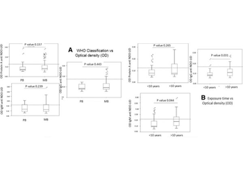
ELISA results using Rabbit Anti-Human IgM. Influence of index case on M. leprae infection levels. Anti-natural octyl disaccharide-leprosy IDRI diagnostic (NDO-LID) antibody levels in children and adolescents were measured by ELISA, using either protein A, anti- IgM or anti-IgG to detect responses. In a, samples were stratified by reported WHO operational classification of the index case as either MB (n = 66) or PB (n = 16). In b, samples were stratified by estimated duration of exposure to the index leprosy case as either less than 10 years (n = 45) or greater than 10 years (n = 37). Data are displayed as box and whisker plots, with the box representing the Q1 to Q3 interquartile range and the horizontal bar representing the median of the optical density of the samples. Individual dots indicate outliers, and p-values are indicated by the lines above each indicated group.
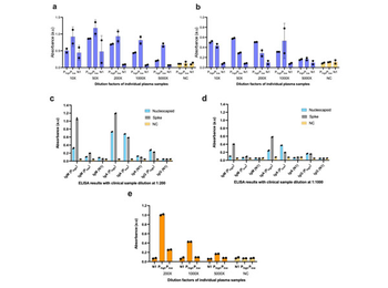
ELISA results using Rabbit Anti-Human IgM. Optimization of the plasma dilutions using clinical plasma samples in a 96- well ELISA format. Serial plasma dilutions were tested in an ELISA plate format with immobilized antigens (a) nucleocapsid and (b) spike protein. Plasma diluted at (c) 200-fold and (d) 1000-fold were compared in ELISA plate formats to detect human IgM, IgA and IgG antibodies against immobilized nucleocapsid and spike protein. Serial plasma dilutions were tested in an ELISA plate format with immobilized S1-RBD to detect human (e) IgM, (f) IgA and (g) IgG. We observed that dilutions between 200X-1000X show a high signal-to-noise ratio in ELISA when detecting IgG against both N and S1 protein antigens (a) and (b), respectively. The dilution factor of 200X (c) detected the less abundant antibody isotypes (IgM and IgA). Similar results were obtained when immobilizing the S1-RBD antigen (e-g), indicating that a dilution factor of 200X was optimal for ELISA experiments. Error bars represent the standard deviation of independent duplicate experiments, biological replicates.
- Item 1 of 4
- Item 1 of 4
- Item 1 of 4
- Item 1 of 4
- Item 1 of 3