You have no items in your shopping cart.
GRP78 Antibody
SKU: orb67383
Featured
Description
Images & Validation
−Item 1 of 4
| Tested Applications | ICC, IF, WB |
|---|---|
| Dilution range | WB (1:2000), ICC/IF (1:100) |
| Reactivity | Bovine, Frog, Fungi, Hamster, Human, Monkey, Mouse, Plant, Rabbit, Rat |
| Application Notes |
Key Properties
−| Host | Mouse |
|---|---|
| Clonality | Monoclonal |
| Isotype | IgG2b |
| Clone No. | 1H11-1H7 |
| Immunogen | His-tagged human GRP78 |
| Target | GRP78 |
| Molecular Weight | 78kDa |
| Purification | Protein G Purified |
Storage & Handling
−| Storage | Maintain refrigerated at 2-8°C for up to 2 weeks. For long term storage store at -20°C in small aliquots to prevent freeze-thaw cycles. |
|---|---|
| Buffer/Preservatives | PBS pH 7.4, 50% glycerol, 0.09% sodium azide *Storage buffer changes when conjugated |
| Concentration | 1 mg/ml |
| Disclaimer | For research use only |
Alternative Names
−GRP78, GRP-78, Grp78, GRP 78, HSPA5, HSPA 5, Heat shock 70 kDa protein 5, Heat Shock 70kDa Protein 5, BiP, BIP, Binding immunoglobulin protein, Immunoglobulin heavy chain-binding protein, Immunoglobulin Heavy Chain Binding Protein, Endoplasmic reticulum lumenal Ca(2+)-binding protein grp78, Endoplasmic reticulum lumenal, Ca2+ binding protein grp78, Glucose Regulated Protein 78kDa, 78 kDa glucose-regulated protein, 78 kDa glucose regulated protein, HSCe70, MIF2, mBiP, Sez7, AL022860, AU019543, D2Wsu141e, D2Wsu17e, FLJ26106, GRP78_HUMAN
Similar Products
−- Item 1 of 8
- Item 1 of 6
- Item 1 of 6
GRP78 BiP/HSPA5 Antibody [orb76328]
ICC, IHC, WB
Human, Mouse, Rat
Rabbit
Polyclonal
Unconjugated
100 μg - Item 1 of 3
GRP78 Rabbit Polyclonal Antibody [orb10751]
IF, IHC-Fr, IHC-P, WB
Mouse, Porcine
Human, Rat
Rabbit
Polyclonal
Unconjugated
50 μl, 100 μl, 200 μl - Item 1 of 7
HSP A5 Polyclonal Antibody [orb1413406]
IF, IHC-P, WB
Human, Mouse, Rat
Rabbit
Polyclonal
Unconjugated
100 μl

Quality Guarantee
Explore bioreagents carefree to elevate your research. All our products are rigorously tested for performance. If a product does not perform as described on its datasheet, our scientific support team will provide expert troubleshooting, a prompt replacement, or a refund. For full details, please see our Terms & Conditions and Buying Guide. Contact us at [email protected].
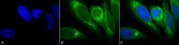
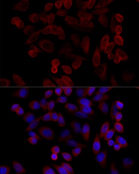
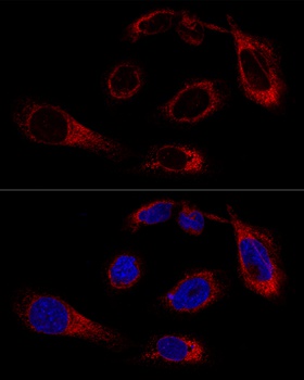
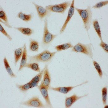
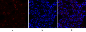
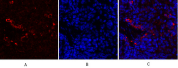
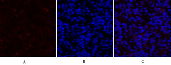
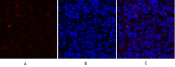
Immunocytochemistry/Immunofluorescence analysis using Mouse Anti-GRP78 Monoclonal Antibody, Clone 1H11-1H7. Tissue: Heat Shocked cervical cancer cells (HeLa). Species: Human. Fixation: 2% Formaldehyde for 20 min at RT. Primary Antibody: Mouse Anti-GRP78 Monoclonal Antibody at 1:100 for 12 hours at 4°C. Secondary Antibody: FITC Goat Anti-Mouse (green) at 1:200 for 2 hours at RT. Counterstain: DAPI (blue) nuclear stain at 1:40000 for 2 hours at RT. Localization: Endoplasmic reticulum lumen. Melanosome. Magnification: 100x. (A) DAPI (blue) nuclear stain. (B) Anti-GRP78 Antibody. (C) Composite. Heat Shocked at 42°C for 1h.
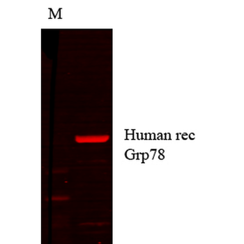
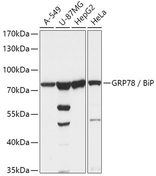
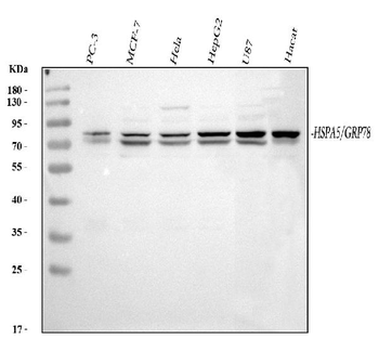
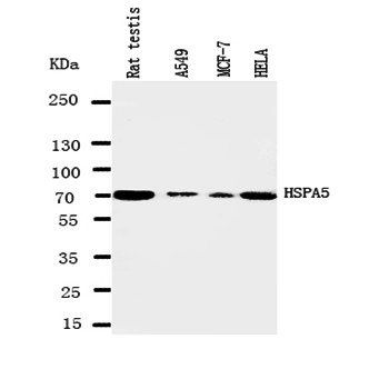
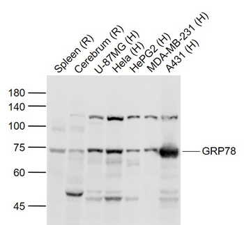
Western Blot analysis of Human cell lysates showing detection of GRP78 protein using Mouse Anti-GRP78 Monoclonal Antibody, Clone 1H11-1H7. Primary Antibody: Mouse Anti-GRP78 Monoclonal Antibody at 1:1000.
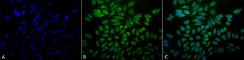
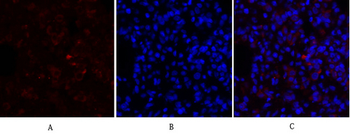
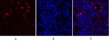
Immunocytochemistry/Immunofluorescence analysis using Mouse Anti-GRP78 Monoclonal Antibody, Clone 1H11-1H7. Tissue: Heat Shocked cervical cancer cells (HeLa). Species: Human. Fixation: 2% Formaldehyde for 20 min at RT. Primary Antibody: Mouse Anti-GRP78 Monoclonal Antibody at 1:100 for 12 hours at 4°C. Secondary Antibody: FITC Goat Anti-Mouse (green) at 1:200 for 2 hours at RT. Counterstain: DAPI (blue) nuclear stain at 1:40000 for 2 hours at RT. Localization: Endoplasmic reticulum lumen. Melanosome. Magnification: 20x. (A) DAPI (blue) nuclear stain. (B) Anti-GRP78 Antibody. (C) Composite. Heat Shocked at 42°C for 1h.
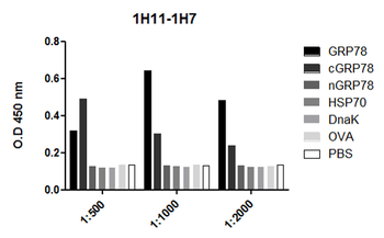
ELISA analysis using Mouse Anti-GRP78 Monoclonal Antibody, Clone 1H11-1H7. Primary Antibody: Mouse Anti-GRP78 Monoclonal Antibody. Secondary Antibody: Goat anti-mouse IgG: HRP at 1:10000.
Quick Database Links
UniProt Details
− No UniProt data available
NCBI Gene Details
− No NCBI Gene data available
NCBI Reference Sequences
−Associated Accession Numbers
Curated reference sequences for the gene transcript and protein product| Protein | NP_001156906.1 |
|---|
Documents Download
Datasheet
Product Information
Request a Document
Protocol Information
WB
Western Blot (IB, immunoblot)
IF
Immunofluorescence
ICC
Immunocytochemistry
GRP78 Antibody (orb67383)

- 0.0
Based on 0 reviews
Participating in our Biorbyt product reviews program enables you to support fellow scientists by sharing your firsthand experience with our products.
Login to Submit a ReviewAvailable Sizes
Select a size below