You have no items in your shopping cart.
Cart summary
Item 1 of 2
Item 1 of 2
Fab Rabbit IgG (H&L) Antibody
Catalog Number: orb348489

| Catalog Number | orb348489 |
|---|---|
| Category | Antibodies |
| Description | Fab Rabbit IgG (H&L) antibody |
| Clonality | Polyclonal |
| Species/Host | Donkey |
| Isotype | IgG Fab |
| Conjugation | Unconjugated |
| Reactivity | Rabbit |
| Form/Appearance | Liquid (sterile filtered) |
| Concentration | 1.0 mg/mL |
| Buffer/Preservatives | 0.01% (w/v) Sodium Azide |
| Purity | This product was prepared from monospecific antiserum by immunoaffinity chromatography using Rabbit IgG coupled to agarose beads followed by solid phase adsorption(s) to remove any unwanted reactivities, papain digestion and chromatographic separation. Assay by immunoelectrophoresis resulted in a single precipitin arc against anti-Donkey Serum. No reaction was observed against anti-Papain or anti-Donkey IgG F(c). |
| Immunogen | Rabbit IgG whole molecule |
| Tested applications | ELISA, IHC, WB |
| Dilution range | ELISA: 1:20,000 - 1:100,000, IHC: 1:1,000 - 1:5,000, WB: 1:2,000 - 1:10,000 |
| Application notes | Fab Anti-Rabbit IgG (H&L) Antibody has been tested by SDS-PAGE and is suitable for highly specific immunological methods requiring extremely low background levels, absence of F(c) mediated binding, lot-to-lot consistency, high titer and specificity. |
| Antibody Type | Secondary Antibody |
| Storage | Store vial at 4° C prior to opening. This product is stable at 4° C as an undiluted liquid. Dilute only prior to immediate use. |
| Alternative names | Donkey Fab Anti-Rabbit IgG Antibody, Donkey Fab Fr Read more... |
| Note | For research use only |
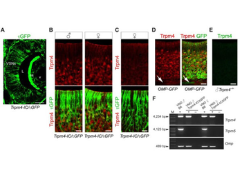
Double labeling of Trpm4 (red) and τGFP (green). Trpm4 is expressed in VSNs of sexually naïve male and female mice. (A) τGFP immunostaining (green) in a coronal cryosection of the left VNO of a 7-week-old Trpm4-IC/eR26-τGFP mouse reports widespread Trpm4 gene expression in sensory neurons and in supporting cells of the VNE. τGFP-IR is also present in cells of the non-sensory (ns) epithelium and in vascular endothelial cells. (B) Magnification of the VNE of male (♂) and female (♀) Trpm4-τGFP reporter mice show that Trpm4 protein (red) colocalizes with τGFP fluorescence (green) in VSNs but is absent in supporting cells. (C) In about 50% of females, VSNs were devoid of Trpm4 protein despite the presence of τGFP. (D) The vast majority of Trpm4+ VSNs (red) colocalizes with the olfactory marker protein (OMP, green), a marker for mature VSNs, in somata, dendrites and dendritic knobs, but not in VSN axon bundles (arrows). (E) The specificity of the Trpm4 antibody is verified by the absence of immunoreactivity in the VNO of Trpm4−/− male mice. (F) RT-PCR analysis of Trpm4 and Trpm5 mRNA prepared from whole VNO and from isolated VSNs (7–10 cells/sample) of male and female B6 and Trpm4-IC/eR26-τGFP mice. Sequence analysis confirmed that the 4.2 kb Trpm4 amplicon (arrowhead) in each sample encodes full-length Trpm4 mRNA. The 4.1 kb Trpm5 amplicon (arrowhead) encoding the full-length Trpm5 mRNA was only detected in whole VNO but absent in isolated VSNs. Identity of dissociated VSNs was verified by RT-PCR for olfactory marker protein (Omp). Control reactions omitting reverse transcriptase (−RT) showed no PCR products ruling out genomic DNA contamination. Scale bars (A) 200 µm, (B-E) 20 µm.
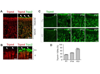
Trpc2 but not Trpm4 protein localizes to VSN microvilli. (A) Double labeling of Trpm4 (red) and Trpc2 (green) depicts co-localization of both TRP channels in VSN somata and dendrites (right panel). The most apical layer (arrowheads) shows robust Trpc2 labeling (green). (B) High-resolution confocal image (1- µm optical section) of the dendritic endings (d) of VSNs shows that microvilli (mv) are heavily labeled for Trpc2 (green, arrowheads) whereas Trpm4-IR (red) was not detected in the microvilli. (C) Representative examples of Trpm4-IR (green) in coronal sections of VNE from B6 mice at postnatal days (P) 1, P7, P14 and P21. Trpm4 protein expression emerges at around P7. Number of Trpm4+ VSNs increases with age. (D) Quantification of Trpm4+ VSNs over developmental time as percentages of the total number of VSNs determined by nuclear Hoechst staining: P7 (33 ± 1%, n = 2 male and 2 female mice, 13 sections, 3–4 sections/mouse); P14 (45 ± 0.2%, n = 2 male and 2 female mice, 15 sections, 3–4 sections/mouse); P21 (71 ± 4.5%, n = 1 male and 2 female mice, 12 sections, 4 sections/mouse). Individual data points represent the averaged cell counts obtained from a single mouse. Data are expressed as means ± SD. Scale bars (A, B) 20 µm, (C) 2 µm.
- Item 1 of 3
- Item 1 of 1
F(ab')2 Bovine IgG (H&L) Antibody Fluorescein Conjugated [orb348075]
DOT, FC, FLISA, IF
Bovine
Rabbit
Polyclonal
FITC
500 μl - Item 1 of 1
F(ab')2 Bovine IgG (H&L) Antibody Texas Red Conjugated [orb348080]
DOT, IF
Bovine
Rabbit
Polyclonal
Texas Red
500 μl - Item 1 of 1
F(ab')2 Chicken IgG (H&L) Antibody Peroxidase Conjugated [orb348093]
ELISA, IHC, WB
Gallus
Rabbit
Polyclonal
HRP
500 μg - Item 1 of 1
F(ab')2 Chicken IgG (H&L) Antibody Texas Red Conjugated [orb348097]
DOT, IF
Gallus
Rabbit
Polyclonal
Texas Red
500 μl