You have no items in your shopping cart.
MCL1 Antibody
SKU: orb215887
Description
Images & Validation
−Item 1 of 5
| Tested Applications | IHC, WB |
|---|---|
| Reactivity | Human, Mouse, Rat |
| Application Notes |
Key Properties
−| Antibody Type | Primary Antibody |
|---|---|
| Host | Rabbit |
| Clonality | Polyclonal |
| Isotype | Rabbit IgG |
| Immunogen | E.coli-derived human MCL1 recombinant protein (Position: M1-R350). Human MCL1 shares 82% and 78% amino acid (aa) sequences identity with mouse and rat MCL1, respectively. |
| Molecular Weight | 37 kDa |
| Purification | Immunogen affinity purified. |
Storage & Handling
−| Storage | Maintain refrigerated at 2-8°C for up to 2 weeks. For long term storage store at -20°C in small aliquots to prevent freeze-thaw cycles. |
|---|---|
| Form/Appearance | Lyophilized |
| Concentration | Adding 0.2 ml of distilled water will yield a concentration of 500 μg/ml. |
| Disclaimer | For research use only |
Alternative Names
−Induced myeloid leukemia cell differentiation protein Mcl-1; Bcl-2-like protein 3; Bcl2-L-3; Bcl-2-related protein EAT/mcl1; mcl1/EAT; MCL1; BCL2L3
Similar Products
−- Item 1 of 6
MCL1 Rabbit Polyclonal Antibody [orb500687]
FC, IF, IHC-Fr, IHC-P, WB
Canine, Equine, Porcine, Rabbit
Human, Mouse, Rat
Rabbit
Polyclonal
Unconjugated
50 μl, 100 μl, 200 μl - Item 1 of 7
Mcl-1 Polyclonal Antibody [orb1411610]
IF, IHC-P, WB
Human, Mouse, Rat
Rabbit
Polyclonal
Unconjugated
100 μl - Item 1 of 1
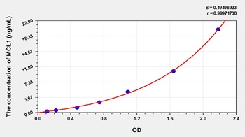
Human Myeloid Cell Leukemia Sequence 1, Bcl2 Related (MCL1) ELISA Kit [orb777797]
Human
0.32-20 ng/mL
0.123 ng/mL
96 T, 48 T - Item 1 of 1
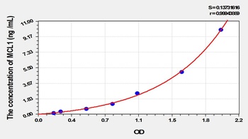
Mouse Myeloid Cell Leukemia Sequence 1, Bcl2 Related (MCL1) ELISA Kit [orb780844]
Mouse
0.16-10 ng/mL
0.062 ng/mL
48 T, 96 T - Item 1 of 4
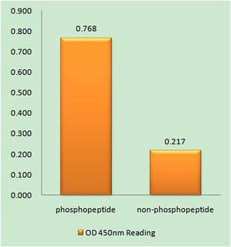
Mcl-1 (phospho Ser159) rabbit pAb [orb769080]
ELISA, IF, WB
Human, Mouse
Polyclonal
Unconjugated
50 μl, 100 μl

Quality Guarantee
Explore bioreagents carefree to elevate your research. All our products are rigorously tested for performance. If a product does not perform as described on its datasheet, our scientific support team will provide expert troubleshooting, a prompt replacement, or a refund. For full details, please see our Terms & Conditions and Buying Guide. Contact us at [email protected].

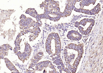
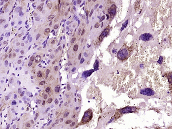
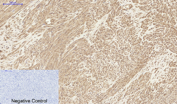
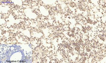
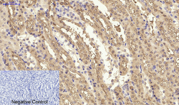
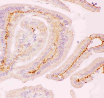
IHC analysis of MCL1 using anti-MCL1 antibody. MCL1 was detected in a paraffin-embedded section of human intestinal cancer tissue. Heat mediated antigen retrieval was performed in EDTA buffer (pH8.0, epitope retrieval solution). The tissue section was blocked with 10% goat serum. The tissue section was then incubated with 1 µg/ml rabbit anti-MCL1 Antibody overnight at 4°C. Biotinylated goat anti-rabbit IgG was used as secondary antibody and incubated for 30 minutes at 37°C. The tissue section was developed using Strepavidin-Biotin-Complex (SABC) with DAB as the chromogen.
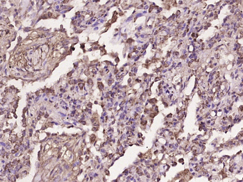
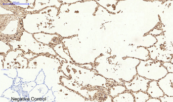
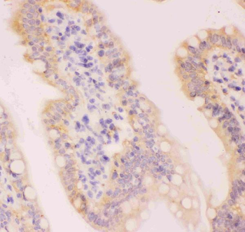
IHC analysis of MCL1 using anti-MCL1 antibody. MCL1 was detected in a paraffin-embedded section of mouse intestine tissue. Heat mediated antigen retrieval was performed in EDTA buffer (pH8.0, epitope retrieval solution). The tissue section was blocked with 10% goat serum. The tissue section was then incubated with 1 µg/ml rabbit anti-MCL1 Antibody overnight at 4°C. Biotinylated goat anti-rabbit IgG was used as secondary antibody and incubated for 30 minutes at 37°C. The tissue section was developed using Strepavidin-Biotin-Complex (SABC) with DAB as the chromogen.
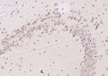
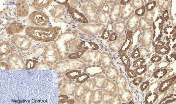
IHC analysis of MCL1 using anti-MCL1 antibody. MCL1 was detected in a paraffin-embedded section of rat intestine tissue. Heat mediated antigen retrieval was performed in EDTA buffer (pH8.0, epitope retrieval solution). The tissue section was blocked with 10% goat serum. The tissue section was then incubated with 1 µg/ml rabbit anti-MCL1 Antibody overnight at 4°C. Biotinylated goat anti-rabbit IgG was used as secondary antibody and incubated for 30 minutes at 37°C. The tissue section was developed using Strepavidin-Biotin-Complex (SABC) with DAB as the chromogen.

Western blot analysis of MCL1 using anti-MCL1 antibody. Electrophoresis was performed on a 5-20% SDS-PAGE gel at 70V (Stacking gel) / 90V (Resolving gel) for 2-3 hours. Lane 1: recombinant human MCL1 protein 0.5 ng. After electrophoresis, proteins were transferred to a nitrocellulose membrane at 150 mA for 50-90 minutes. Blocked the membrane with 5% non-fat milk/TBS for 1.5 hour at RT. The membrane was incubated with rabbit anti-MCL1 antigen affinity purified polyclonal antibody at 0.5 µg/mL overnight at 4°C, then washed with TBS-0.1% Tween 3 times with 5 minutes each and probed with a goat anti-rabbit IgG-HRP secondary antibody at a dilution of 1:5000 for 1.5 hour at RT. The signal is developed using an Enhanced Chemiluminescent detection (ECL) kit with Tanon 5200 system. A specific band was detected for MCL1 at approximately 40 kDa. The expected band size for MCL1 is at 40 kDa.
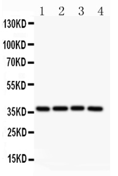
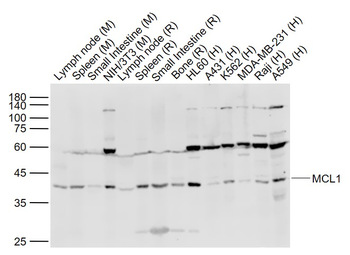
Western blot analysis of MCL1 using anti-MCL1 antibody. Electrophoresis was performed on a 5-20% SDS-PAGE gel at 70V (Stacking gel) / 90V (Resolving gel) for 2-3 hours. The sample well of each lane was loaded with 30 ug of sample under reducing conditions. Lane 1: rat spleen tissue lysates, Lane 2: human HepG2 whole cell lysates, Lane 3: human MCF-7 whole cell lysates, Lane 4: human COLO320 whole cell lysates. After electrophoresis, proteins were transferred to a nitrocellulose membrane at 150 mA for 50-90 minutes. Blocked the membrane with 5% non-fat milk/TBS for 1.5 hour at RT. The membrane was incubated with rabbit anti-MCL1 antigen affinity purified polyclonal antibody at 0.5 µg/mL overnight at 4°C, then washed with TBS-0.1% Tween 3 times with 5 minutes each and probed with a goat anti-rabbit IgG-HRP secondary antibody at a dilution of 1:5000 for 1.5 hour at RT. The signal is developed using an Enhanced Chemiluminescent detection (ECL) kit with Tanon 5200 system. A specific band was detected for MCL1 at approximately 37 kDa. The expected band size for MCL1 is at 37 kDa.
Quick Database Links
UniProt
UniProt Details
− No UniProt data available
Documents Download
Datasheet
Product Information
Request a Document
Protocol Information
WB
Western Blot (IB, immunoblot)
IHC
Immunohistochemistry
MCL1 Antibody (orb215887)

- 0.0
Based on 0 reviews
Participating in our Biorbyt product reviews program enables you to support fellow scientists by sharing your firsthand experience with our products.
Login to Submit a ReviewAvailable Sizes
Select a size below