You have no items in your shopping cart.
Cart summary
Item 1 of 3
Item 1 of 3
Lamin A/C Antibody
Catalog Number: orb1474439

Product Properties
| Catalog Number | orb1474439 |
|---|---|
| Category | Antibodies |
| Description | The Lamin A/C Antibody is suitable for IF, IHC, WB. It is a Monoclonal, Unconjugated antibody which raised against A synthetic peptide of human Lamin A/C .Purification: The antibody was purified by immunogen affinity chromatography. |
| Clonality | Monoclonal |
| Species/Host | Rabbit |
| Conjugation | Unconjugated |
| Reactivity | Human, Mouse, Rat |
| Form/Appearance | Liquid in 50mM Tris-Glycine (pH 7.4), 0.15M NaCl, 50% Glycerol, 0.01% Sodium azide and 0.05% rAlbumin. |
| Purification | The antibody was purified by immunogen affinity chromatography. |
| Immunogen | A synthetic peptide of human Lamin A/C |
| UniProt ID | P48678, P02545 |
| Tested applications | IF, IHC, WB |
| Dilution range | WB (1/500 - 1/1000), IH (1/50 - 1/100), IF/IC (1/50 - 1/100) |
| Antibody Type | Primary Antibody |
| Storage | Maintain refrigerated at 2-8°C for up to 2 weeks. For long term storage store at -20°C in small aliquots to prevent freeze-thaw cycles. |
| Alternative names | LMN1; Prelamin-A/C |
| Note | For research use only |
| Entrez | 16905, 4000 |
| Expiration Date | 12 months from date of receipt. |
Images
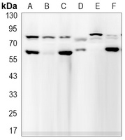
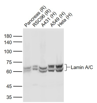
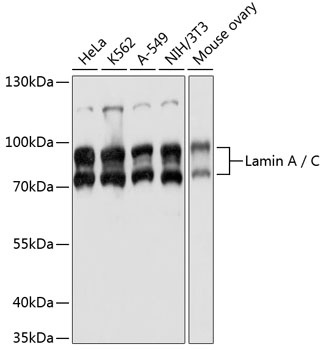
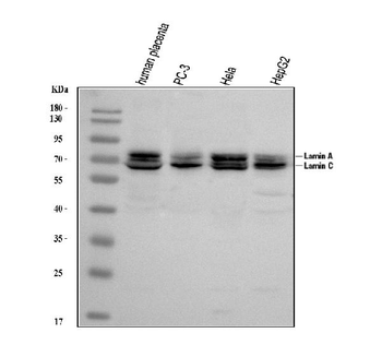
Western blot analysis of Lamin A/C expression in NIH3T3 (A), C2C12 (B), U251 (C), Lncap (D), A549 (E) whole cell lysates. (Predicted band size: 74 kD; Observed band size: 74, 63 kD)
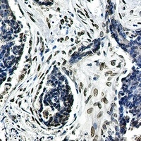
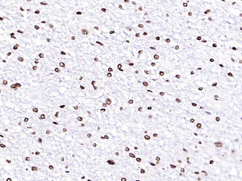
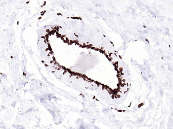
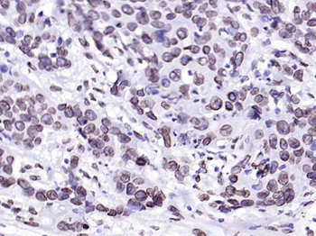
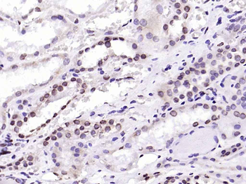
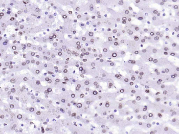
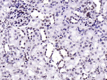
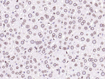
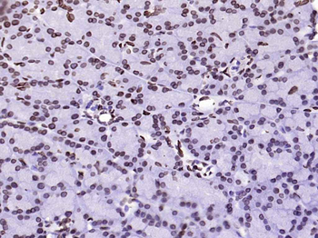
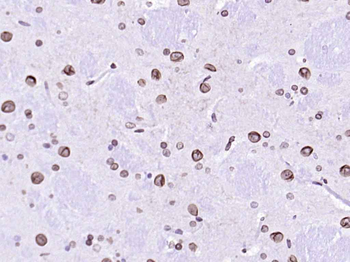
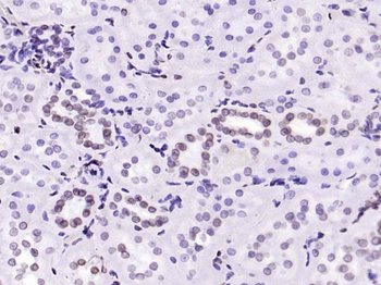
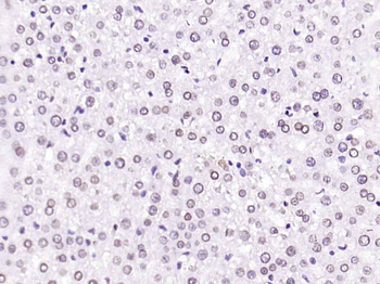
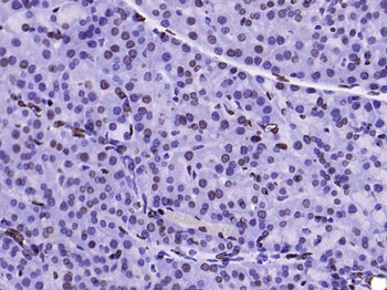
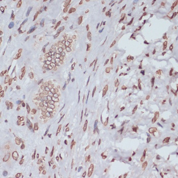
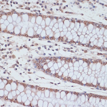
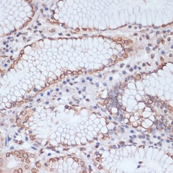
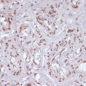
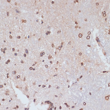
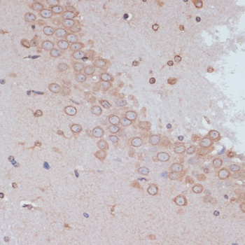
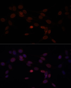
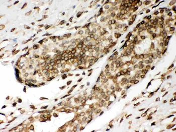
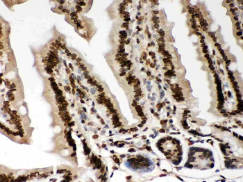
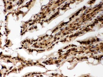
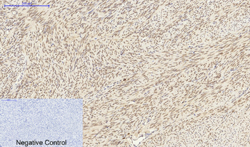
Immunohistochemical analysis of Lamin A/C staining in human breast carcinoma formalin fixed paraffin embedded tissue section. The section was pre-treated using heat mediated antigen retrieval with sodium citrate buffer (pH 6.23). The section was then incubated with the antibody at room temperature and detected using an HRP conjugated compact polymer system. DAB was used as the chromogen. The section was then counterstained with haematoxylin and mounted with DPX.
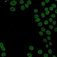
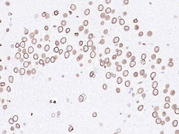
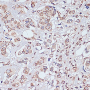
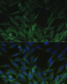
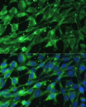
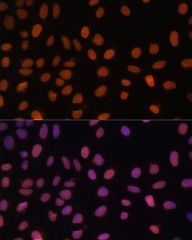
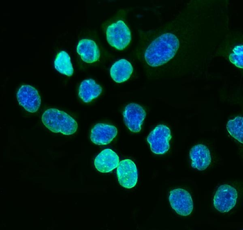
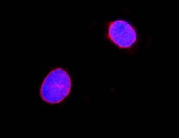
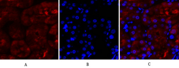
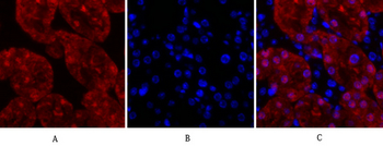
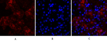
Immunofluorescent analysis of Lamin A/C staining in HeLa cells. Formalin-fixed cells were permeabilized with 0.1% Triton X-100 in TBS for 5-10 minutes and blocked with 3% BSA-PBS for 30 minutes at room temperature. Cells were probed with the primary antibody in 3% BSA-PBS and incubated overnight at 4 °C in a hidified chamber. Cells were washed with PBST and incubated with a AF488-conjugated secondary antibody (green) in PBS at room temperature in the dark.
Similar Products
- Item 1 of 15
Lamin A/C Mouse Monoclonal Antibody [orb783433]
ELISA, ICC, IF, IHC-Fr, IHC-P, WB
Bovine, Canine, Equine, Porcine
Human, Mouse, Rat
Mouse
Monoclonal
Unconjugated
100 μl, 50 μl - Item 1 of 12
- Item 1 of 7
Lamin A+C/LMNA Antibody [orb234326]
FC, ICC, IF, IHC, WB
Human, Mouse, Rat
Rabbit
Polyclonal
Unconjugated
100 μg - Item 1 of 7
Lamin A/C Polyclonal Antibody [orb1413238]
IF, IHC-P, WB
Human, Mouse, Rat
Rabbit
Polyclonal
Unconjugated
100 μl - Item 1 of 1


Reviews
Lamin A/C Antibody (orb1474439)

- 0.0
Based on 0 reviews
Participating in our Biorbyt product reviews program enables you to support fellow scientists by sharing your firsthand experience with our products.
Login to Submit a Review