You have no items in your shopping cart.
Cart summary
Item 1 of 9
Item 1 of 9
VPS35 Antibody: HRP
Catalog Number: orb612803

Product Properties
| Catalog Number | orb612803 |
|---|---|
| Category | Antibodies |
| Description | Mouse monoclonal antibody against VPS35 conjugated to HRP |
| Target | VPS35 |
| Clonality | Monoclonal |
| Species/Host | Mouse |
| Isotype | IgG1 |
| Conjugation | HRP |
| Reactivity | Human, Mouse, Rat |
| Concentration | 1 mg/ml |
| Buffer/Preservatives | 73.64mM Carbonate, 54.55mM Ethanolamine, 45.45mM Cyanoborohydride, 18.18mM Sodium Hydroxide and 0.23mM Citrate in dH2O |
| Purification | Protein G Purified |
| Immunogen | Full length recombinant human VSP35 |
| UniProt ID | Q96QK1 |
| MW | 92 kDa |
| Tested applications | ELISA, IHC, WB |
| Dilution range | WB (1:1000); ICC/IF (1:200); IP (1:200); IHC (1:100) |
| Application notes | A 1:1000 dilution of SMC-604 was sufficient for detection of VPS35 in 10 µg of SH-SY5Y by ECL immunoblot analysis using Goat Anti-Mouse IgG:HRP as the secondary antibody. |
| Clone Number | 8A3 |
| Storage | Conjugated antibodies should be stored according to the product label |
| Alternative names | Vacuolar protein sorting-associated protein 35, ME Read more... |
| Research Area | Cell Biology, Neuroscience, Protein Biochemistry, Read more... |
| Note | For research use only |
| Entrez | 55737 |
| NCBI | NP_060676.2 |
| Expiration Date | 12 months from date of receipt. |
Images
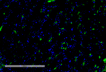
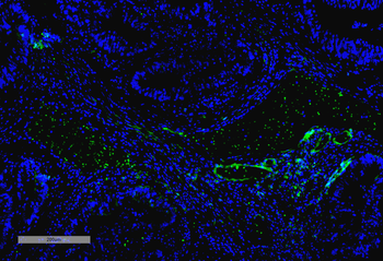
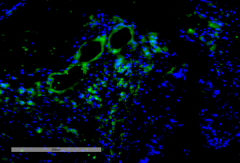
Immunohistochemistry analysis using Mouse Anti-VPS35 Monoclonal Antibody, Clone 8A3. Tissue: Kidney. Species: Mouse. Primary Antibody: Mouse Anti-VPS35 Monoclonal Antibody at 1:100 for Overnight at 4°C, then 30 min at 37°C. Secondary Antibody: Goat Anti-Mouse IgG (H+L): FITC for 45 min at 37°C. Counterstain: DAPI for 3 min at RT. Magnification: 20X.
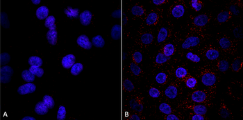
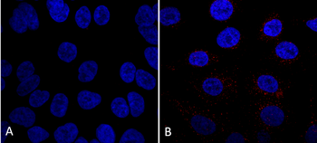
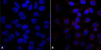
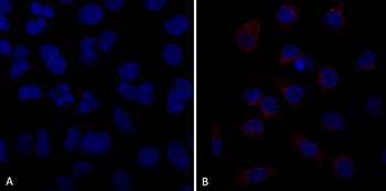
Immunocytochemistry/Immunofluorescence analysis using Mouse Anti-VPS35 Monoclonal Antibody, Clone 8A3. Tissue: A549 cells. Species: Human. Primary Antibody: Mouse Anti-VPS35 Monoclonal Antibody at 1:5 (tissue culture supernatant). Secondary Antibody: Donkey anti-mouse: Alexa Fluor 594 at 1:4000 in 0.2% BSA PBS. Counterstain: DAPI. Localization: Vesicles. A) VPS35 KO A549 cells B) WT A549 cells.
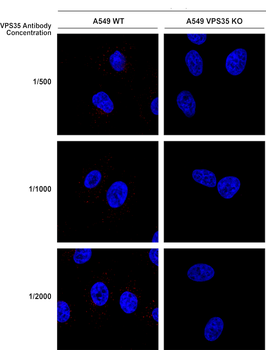
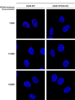
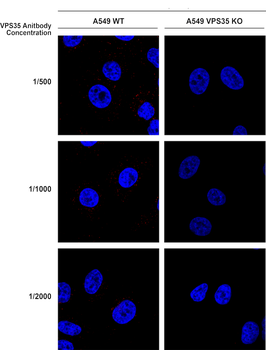
Immunocytochemistry/Immunofluorescence analysis using Mouse Anti-VPS35 Monoclonal Antibody, Clone 8A3. Tissue: A549 WT, VPS35 KO cells. Species: Human. Primary Antibody: Mouse Anti-VPS35 Monoclonal Antibody. Secondary Antibody: Donkey Anti-Mouse AlexaFluor 594. Clone can detect VPS35 at 1/2000 concentration.
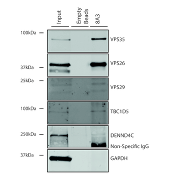
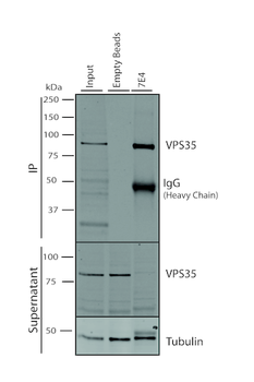
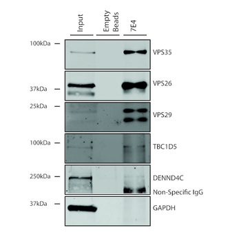
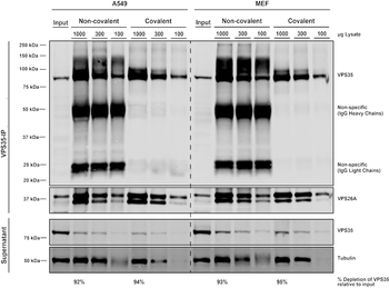
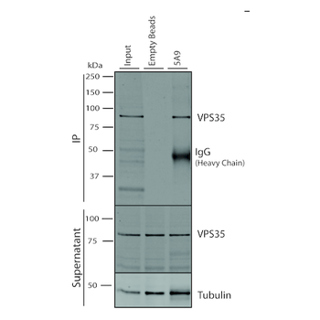
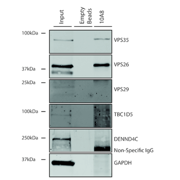
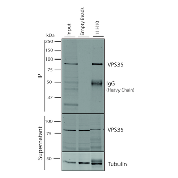
Immunoprecipitation analysis using Mouse Anti-VPS35 Monoclonal Antibody, Clone 8A3. Tissue: A549 cells. Species: Human. Primary Antibody: Mouse Anti-VPS35 Monoclonal Antibody.
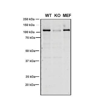
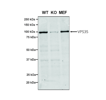
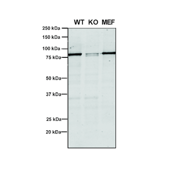
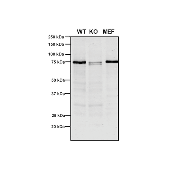
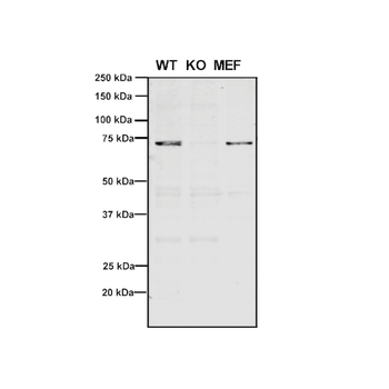
Western Blot analysis of Human, Mouse A549, MEF showing detection of VPS35 protein using Mouse Anti-VPS35 Monoclonal Antibody, Clone 8A3. Lane 1: Molecular Weight Ladder. Lane 2: VPS35 KO A549 cells. Lane 3: mouse embryonic fibroblast cells. Load: 8 μg each A549 and MEF. Primary Antibody: Mouse Anti-VPS35 Monoclonal Antibody at 1:5 (tissue culture supernatant). Secondary Antibody: Donkey anti-mouse IRDye 800CW at 1:25000 in TBS-T.
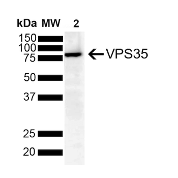
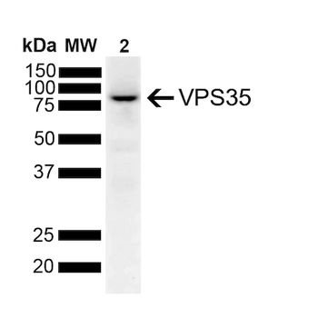
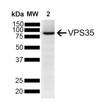
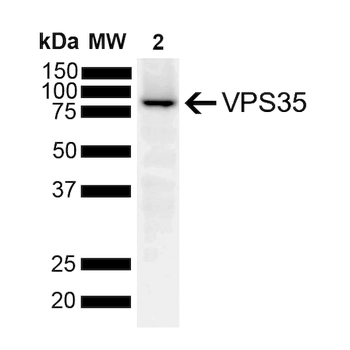
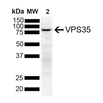
Western Blot analysis of Human SH-SY5Y showing detection of VPS35 protein using Mouse Anti-VPS35 Monoclonal Antibody, Clone 8A3. Lane 1: Molecular Weight Ladder. Lane 2: SH-SY5Y (10 ug). Load: 10 μg. Block: 5% Skim Milk powder in TBST. Primary Antibody: Mouse Anti-VPS35 Monoclonal Antibody at 1:1000 for 2 hours at RT with shaking. Secondary Antibody: Goat anti-mouse IgG:HRP at 1:4000 for 1 hour at RT with shaking. Color Development: Chemiluminescent for HRP (Moss) for 5 min in RT.
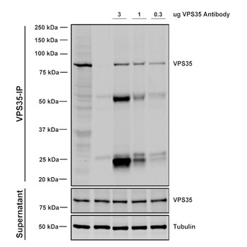
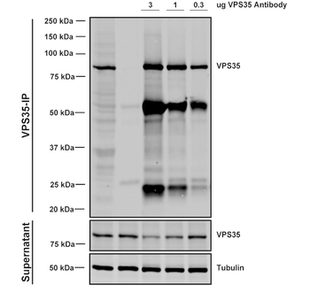
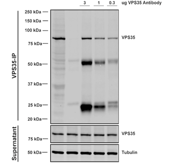
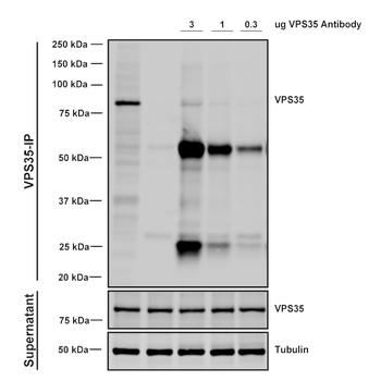
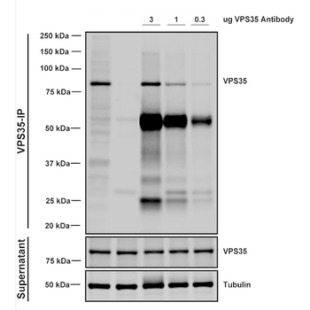
Immunoprecipitation analysis using Mouse Anti-VPS35 Monoclonal Antibody, Clone 8A3. Tissue: A549 cells. Species: Human. Primary Antibody: Mouse Anti-VPS35 Monoclonal Antibody. Three amounts of (3, 1 and 0.3 ug) were non-covalently coupled to 10uL of A/G sepharose beads for 1 hour at 4°C and next incubated with 250ug of A549 lysate for 2 hours at 4°C.

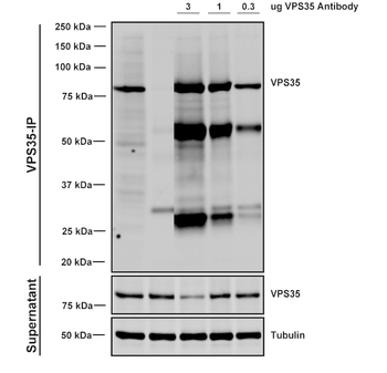
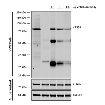
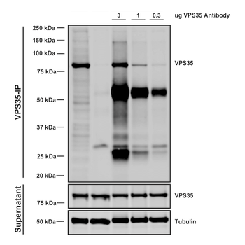
Immunoprecipitation analysis using Mouse Anti-VPS35 Monoclonal Antibody, Clone 8A3. Tissue: embryonic fibroblast. Species: Mouse. Primary Antibody: Mouse Anti-VPS35 Monoclonal Antibody. Three amounts of (3, 1 and 0.3 ug) were non-covalently coupled to 10uL of A/G sepharose beads for 1 hour at 4°C and next incubated with 250ug of MEF lysate for 2 hours at 4°C.
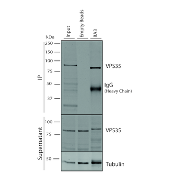
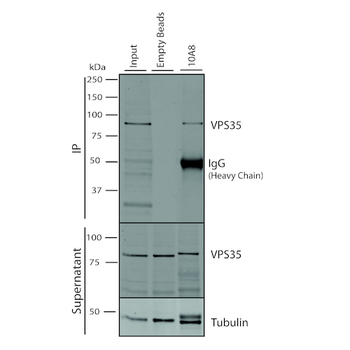
Immunoprecipitation analysis using Mouse Anti-VPS35 Monoclonal Antibody, Clone 8A3. Tissue: A549 cells. Species: Human. Primary Antibody: Mouse Anti-VPS35 Monoclonal Antibody. 500 μL cell culture supernatants were incubated with 10 μL of Protein A/G resin beads for 1 hour at 4°C.
Similar Products
- Item 1 of 8
- Item 1 of 8
- Item 1 of 8
- Item 1 of 7

VPS35 Rabbit Polyclonal Antibody (HRP) [orb475776]
IHC-Fr, IHC-P, WB
Bovine, Canine, Equine, Gallus, Porcine, Rabbit, Sheep
Human, Mouse, Rat
Rabbit
Polyclonal
HRP
100 μl


Reviews
VPS35 Antibody: HRP (orb612803)

- 0.0
Based on 0 reviews
Participating in our Biorbyt product reviews program enables you to support fellow scientists by sharing your firsthand experience with our products.
Login to Submit a Review