You have no items in your shopping cart.
Cart summary
Item 1 of 7
Item 1 of 7
Tyrosine Hydroxylase Recombinant Rabbit Monoclonal Antibody
Catalog Number: orb1499388

Product Properties
| Catalog Number | orb1499388 |
|---|---|
| Category | Antibodies |
| Description | Tyrosine Hydroxylase Recombinant Rabbit Monoclonal Antibody |
| Target | TH |
| Clonality | Recombinant |
| Species/Host | Rabbit |
| Isotype | IgG |
| Conjugation | Unconjugated |
| Reactivity | Human, Mouse, Rat |
| Predicted Reactivity | Mouse, Rat |
| Form/Appearance | Liquid |
| Concentration | 1mg/ml |
| Buffer/Preservatives | 0.01M TBS (pH7.4) with 1% rAlbumin, 0.02% Proclin300 and 50% Glycerol. |
| Purification | Affinity purified by Protein A |
| Immunogen | A synthesized peptide derived from human Tyrosine hydroxylase (500-528aa) |
| UniProt ID | P07101 |
| MW | 60 kDa |
| Tested applications | FC, ICC, IF, IHC-Fr, IHC-P, WB |
| Dilution range | WB=1:500-2000, IHC-P=1:100-500, IHC-F=1:100-500, ICC/IF=1:50-200, IF=1:100-500, Flow-Cyt=1:50-100 |
| Antibody Type | Primary Antibody |
| Clone Number | 8B6 |
| Storage | Maintain refrigerated at 2-8°C for up to 2 weeks. For long term storage store at -20°C in small aliquots to prevent freeze-thaw cycles. |
| Alternative names | DYT14; DYT5b; ple; Protein Pale; c; The; TYH; Tyro Read more... |
| Research Area | Dopamine, Hormone Biosynthesis, Hypoxia, Neuroscie Read more... |
| Note | For research use only |
| Expiration Date | 12 months from date of receipt. |
Images
ICC staining of Tyrosine Hydroxylase in N2A cells (green). Formalin fixed cells were permeabilized with 0.1% Triton X-100 in TBS for 10 minutes at room temperature and blocked with 1% Blocker BSA for 15 minutes at room temperature. Cells were probed with the primary antibody (orb1499388, 1/50) for 1 hour at room temperature, washed with PBS. Alexa Fluor®488 Goat anti-Rabbit IgG was used as the secondary antibody at 1/1000 dilution. The nuclear counter stain is DAPI (blue).

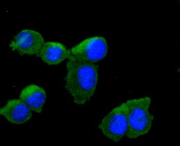
ICC staining of Tyrosine Hydroxylase in NIH/3T3 cells (green). Formalin fixed cells were permeabilized with 0.1% Triton X-100 in TBS for 10 minutes at room temperature and blocked with 1% Blocker BSA for 15 minutes at room temperature. Cells were probed with the primary antibody (orb1499388, 1/50) for 1 hour at room temperature, washed with PBS. Alexa Fluor®488 Goat anti-Rabbit IgG was used as the secondary antibody at 1/1000 dilution. The nuclear counter stain is DAPI (blue).
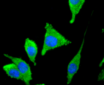
ICC staining of Tyrosine Hydroxylase in SH-SY5Y cells (green). Formalin fixed cells were permeabilized with 0.1% Triton X-100 in TBS for 10 minutes at room temperature and blocked with 1% Blocker BSA for 15 minutes at room temperature. Cells were probed with the primary antibody (orb1499388, 1/50) for 1 hour at room temperature, washed with PBS. Alexa Fluor®488 Goat anti-Rabbit IgG was used as the secondary antibody at 1/1000 dilution. The nuclear counter stain is DAPI (blue).
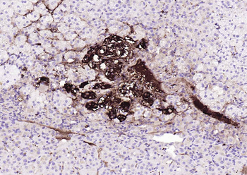
Paraformaldehyde-fixed, paraffin embedded (human adrenal gland), Antigen retrieval by boiling in EDTA buffer buffer (pH9.0) for 15 min, Block endogenous peroxidase by 3% hydrogen peroxide for 20 minutes, Blocking buffer (normal goat serum) at 37°C for 30 min, Incubation with (Tyrosine Hydroxylase) Monoclonal Antibody, Unconjugated (orb1499388) at 1:200 overnight at 4°C, followed by operating according to SP Kit (Rabbit) instructionsand DAB staining.
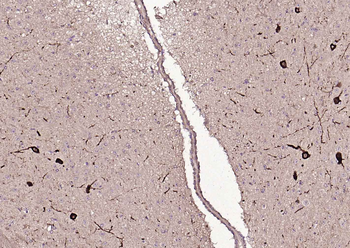
Paraformaldehyde-fixed, paraffin embedded (mouse brain), Antigen retrieval by boiling in EDTA buffer buffer (pH9.0) for 15 min, Block endogenous peroxidase by 3% hydrogen peroxide for 20 minutes, Blocking buffer (normal goat serum) at 37°C for 30 min, Incubation with (Tyrosine Hydroxylase) Monoclonal Antibody, Unconjugated (orb1499388) at 1:200 overnight at 4°C, followed by operating according to SP Kit (Rabbit) instructionsand DAB staining.
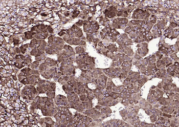
Paraformaldehyde-fixed, paraffin embedded (rat adrenal gland), Antigen retrieval by boiling in EDTA buffer buffer (pH9.0) for 15 min, Block endogenous peroxidase by 3% hydrogen peroxide for 20 minutes, Blocking buffer (normal goat serum) at 37°C for 30 min, Incubation with (Tyrosine Hydroxylase) Monoclonal Antibody, Unconjugated (orb1499388) at 1:200 overnight at 4°C, followed by operating according to SP Kit (Rabbit) instructionsand DAB staining.
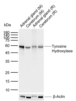
Sample: Lane 1: Mouse Adrenal gland tissue lysates, Lane 2: Mouse Cerebrum tissue lysates, Lane 3: Rat Adrenal gland tissue lysates, Lane 4: Rat Cerebrum tissue lysates, Primary: Anti-Tyrosine Hydroxylase (orb1499388) at 1/1000 dilution, Secondary: IRDye800CW Goat Anti-Rabbit IgG at 1/20000 dilution, Predicted band size: 60 kDa, Observed band size: 60 kDa.
Similar Products
- Item 1 of 1
Rabbit anti-Tyrosine Hydroxylase Recombinant Monoclonal Antibody [orb1519607]
ICC, IHC, WB
Human, Mouse
Rabbit
Recombinant
Unconjugated
10 μl - Item 1 of 1
Rabbit anti-Tyrosine Hydroxylase Recombinant Monoclonal Antibody [orb1519609]
ICC, IHC, WB
Human, Mouse
Rabbit
Recombinant
Unconjugated
100 μl 
Rabbit anti-Tyrosine Hydroxylase Recombinant Monoclonal Antibody [orb1519608]
ICC, IHC, WB
Human, Mouse
Rabbit
Recombinant
Unconjugated
100 μgRecombinant Tyrosine Hydroxylase Rabbit mAb Antibody [orb2988826]
IF, IHC, WB
Human, Mouse, Rat
Rabbit
Monoclonal
Unconjugated
30 μl, 50 μl, 100 μl, 200 μlRecombinant Tyrosine Hydroxylase Rabbit mAb Antibody [orb2989244]
IF, IHC, WB
Human, Mouse, Rat
Rabbit
Monoclonal
Unconjugated
30 μl, 50 μl, 100 μl, 200 μl


Reviews
Tyrosine Hydroxylase Recombinant Rabbit Monoclonal Antibody (orb1499388)

- 0.0
Based on 0 reviews
Participating in our Biorbyt product reviews program enables you to support fellow scientists by sharing your firsthand experience with our products.
Login to Submit a Review