You have no items in your shopping cart.
Cart summary
Item 1 of 3
Item 1 of 3
TrpC5 Antibody: APC
Catalog Number: orb148874

| Catalog Number | orb148874 |
|---|---|
| Category | Antibodies |
| Description | Mouse monoclonal to TrpC5 (APC). Transient receptor potential cation channel, subfamily C, member 5, also known as TRPC5, is a subtype of the TRPC family of mammalian transient receptor potential ion channels. Homo-multimeric TRPC5 and hetero-multimeric TRPC5-TRPC1 channels are activated by extracellular reduced thioredoxin. This activation probably plays a role in rheumatoid arthritis. It has also been recently found to be involved in the action on anaesthetics such as chloroform, halothane and propofol.. |
| Species/Host | Mouse |
| Clonality | Monoclonal |
| Clone Number | N67/15 (Formerly sold as S67-15) |
| Tested applications | ICC, IF, IHC |
| Reactivity | Human, Mouse, Rat |
| Isotype | IgG2b |
| Immunogen | Synthetic peptide amino acids 827-845 of human TrpC5 (also known as short transient receptor potential channel 5, and Htrp5) |
| Concentration | 1 mg/ml |
| Dilution range | WB (1:1000), IHC (1:1000), ICC/IF (1:100) |
| Conjugation | APC |
| MW | 110kDa |
| Target | TRPC5 |
| Entrez | 7224 |
| UniProt ID | Q9UL62 |
| NCBI | NP_036603.1 |
| Storage | Conjugated antibodies should be stored according to the product label |
| Buffer/Preservatives | 95.64mM Phosphate, 2.48mM MES and 2mM EDTA |
| Alternative names | Htrp5 antibody, Short transient receptor potential Read more... |
| Note | For research use only |
| Application notes | 1 µg/ml of SMC-344 was sufficient for detection of TrpC5 in 20 µg of rat brain lysate by colorimetric immunoblot analysis using Goat anti-mouse IgG:HRP as the secondary antibody. |
| Expiration Date | 12 months from date of receipt. |
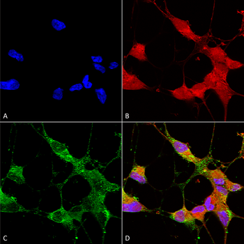
Immunocytochemistry/Immunofluorescence analysis using Mouse Anti-TrpC5 Monoclonal Antibody, Clone N67/15. Tissue: Neuroblastoma cells (SH-SY5Y). Species: Human. Fixation: 4% PFA for 15 min. Primary Antibody: Mouse Anti-TrpC5 Monoclonal Antibody at 1:50 for overnight at 4°C with slow rocking. Secondary Antibody: AlexaFluor 488 at 1:1000 for 1 hour at RT. Counterstain: Phalloidin-iFluor 647 (red) F-Actin stain; Hoechst (blue) nuclear stain at 1:800, 1.6mM for 20 min at RT. (A) Hoechst (blue) nuclear stain. (B) Phalloidin-iFluor 647 (red) F-Actin stain. (C) TrpC5 Antibody (D) Composite.

Immunohistochemistry analysis using Mouse Anti-TrpC5 Monoclonal Antibody, Clone N67/15. Tissue: Brain Slice. Species: Mouse. Fixation: 10% Formalin Solution for 12-24 hours at RT. Primary Antibody: Mouse Anti-TrpC5 Monoclonal Antibody at 1:1000 for 1 hour at RT. Secondary Antibody: HRP/DAB Detection System: Biotinylated Goat Anti-Mouse, Streptavidin Peroxidase, DAB Chromogen (brown) for 30 minutes at RT. Counterstain: Mayer Hematoxylin (purple/blue) nuclear stain at 250-500 μl for 5 minutes at RT. Localization: Nuclear staining. Magnification: 10X.
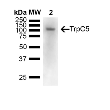
Western Blot analysis of Mouse brain showing detection of 110 kDa TrpC5 protein using Mouse Anti-TrpC5 Monoclonal Antibody, Clone N67/15. Lane 1: Molecular Weight Ladder (MW). Lane 2: Mouse Brain. Load: 15 ug. Block: 5% Skim Milk powder in TBST. Primary Antibody: Mouse Anti-TrpC5 Monoclonal Antibody at 1:1000 for Overnight at 4°C. Secondary Antibody: Goat anti-mouse IgG:HRP at 1:7000 for 1 hour at RT with shaking. Color Development: Chemiluminescent for HRP (Moss) for 5 min in RT. Predicted/Observed Size: 110 kDa.
