You have no items in your shopping cart.
Cart summary
Item 1 of 3
Item 1 of 3
TAK1/MAP3K7 Recombinant Rabbit Monoclonal Antibody
Catalog Number: orb1499366

Product Properties
| Catalog Number | orb1499366 |
|---|---|
| Category | Antibodies |
| Description | TAK1/MAP3K7 Recombinant Rabbit Monoclonal Antibody |
| Target | MAP3K7 |
| Clonality | Recombinant |
| Species/Host | Rabbit |
| Isotype | IgG |
| Conjugation | Unconjugated |
| Reactivity | Human, Mouse |
| Predicted Reactivity | Human, Mouse |
| Form/Appearance | Liquid |
| Concentration | 1mg/ml |
| Buffer/Preservatives | 0.01M TBS (pH7.4) with 1% rAlbumin, 0.02% Proclin300 and 50% Glycerol. |
| Purification | Affinity purified by Protein A |
| Immunogen | KLH conjugated synthetic peptide derived from human TAK1/MAP3K7 |
| UniProt ID | O43318 |
| MW | 67 kDa |
| Tested applications | ICC, WB |
| Dilution range | WB=1:500-1000, ICC/IF=1:50-100 |
| Antibody Type | Primary Antibody |
| Storage | Maintain refrigerated at 2-8°C for up to 2 weeks. For long term storage store at -20°C in small aliquots to prevent freeze-thaw cycles. |
| Alternative names | Mitogen-activated protein kinase kinase kinase 7; Read more... |
| Research Area | Cancer Research, Cell Biology, Inflammation, Infla Read more... |
| Note | For research use only |
| Expiration Date | 12 months from date of receipt. |
Images
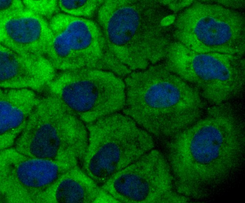
ICC staining of TAK1 in A431 cells (green). Formalin fixed cells were permeabilized with 0.1% Triton X-100 in TBS for 10 minutes at room temperature and blocked with 1% Blocker BSA for 15 minutes at room temperature. Cells were probed with the primary antibody (orb1499366, 1/50) for 1 hour at room temperature, washed with PBS. Alexa Fluor®488 Goat anti-Rabbit IgG was used as the secondary antibody at 1/1000 dilution. The nuclear counter stain is DAPI (blue).
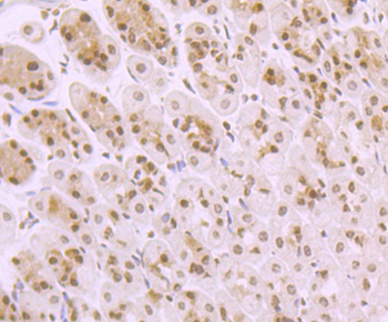
ICC staining of TAK1 in N2A cells (green). Formalin fixed cells were permeabilized with 0.1% Triton X-100 in TBS for 10 minutes at room temperature and blocked with 1% Blocker BSA for 15 minutes at room temperature. Cells were probed with the primary antibody (orb1499366, 1/50) for 1 hour at room temperature, washed with PBS. Alexa Fluor®488 Goat anti-Rabbit IgG was used as the secondary antibody at 1/1000 dilution. The nuclear counter stain is DAPI (blue).
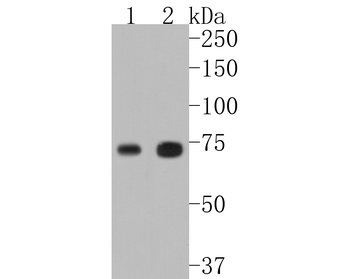
Western blot analysis of TAK1 on different lysates. Proteins were transferred to a PVDF membrane and blocked with 5% BSA in PBS for 1 hour at room temperature. The primary antibody (orb1499366, 1/500) was used in 5% BSA at room temperature for 2 hours. Goat Anti-Rabbit IgG - HRP Secondary Antibody (HA1001) at 1:5000 dilution was used for 1 hour at room temperature. Positive control: Lane 1: MCF-7 cell lysate, Lane 2: A431 cell lysate.
Reviews
TAK1/MAP3K7 Recombinant Rabbit Monoclonal Antibody (orb1499366)

- 0.0
Based on 0 reviews
Participating in our Biorbyt product reviews program enables you to support fellow scientists by sharing your firsthand experience with our products.
Login to Submit a Review