You have no items in your shopping cart.
Cart summary
Item 1 of 5
Item 1 of 5
SP1 Antibody (C-term P692)
Catalog Number: orb1938187

| Catalog Number | orb1938187 |
|---|---|
| Category | Antibodies |
| Description | Affinity Purified Rabbit Polyclonal Antibody (Pab) |
| Species/Host | Rabbit |
| Clonality | Polyclonal |
| Clone Number | RB17350 |
| Tested applications | FC, IF, IHC-P, WB |
| Predicted Reactivity | Mouse, Rat |
| Reactivity | Human |
| Isotype | Rabbit IgG |
| Antibody Type | Primary Antibody |
| Dilution range | IF: 1:10~50, WB: 1:500, WB: 1:1000, IHC-P: 1:50~100, FC: 1:10~50 |
| Form/Appearance | Purified polyclonal antibody supplied in PBS with 0.09% (W/V) sodium azide. This antibody is purified through a protein A column, followed by peptide affinity purification. |
| Conjugation | Unconjugated |
| MW | 80693 Da |
| Target | This SP1 antibody is generated from rabbits immunized with a KLH conjugated synthetic peptide between 677-707 amino acids from the C-terminal region of human SP1. |
| UniProt ID | P08047 |
| NCBI | NP_612482.2, NP_001238754.1, NP_003100.1 |
| Storage | Maintain refrigerated at 2-8°C for up to 2 weeks. For long term storage store at -20°C in small aliquots to prevent freeze-thaw cycles |
| Alternative names | Transcription factor Sp1, SP1, TSFP1 Read more... |
| Note | For research use only |
| Expiration Date | 12 months from date of receipt. |
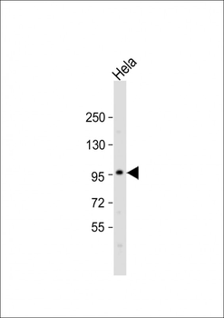
Anti-SP1 Antibody (C-term P692) at 1:500 dilution + Hela whole cell lysate.Lysates/proteins at 20 µg per lane. Secondary Goat Anti-Rabbit IgG, (H+L), Peroxidase conjugated at 1/10000 dilution. Predicted band size: 81 kDa. Blocking/Dilution buffer: 5% NFDM/TBST.
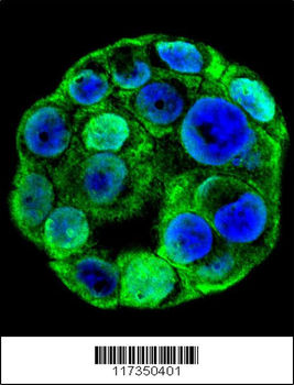
Confocal immunofluorescent analysis of SP1 Antibody (C-term P692) with WiDr cell followed by Alexa Fluor 488-conjugated goat anti-rabbit lgG (green). DAPI was used to stain the cell nuclear (blue).
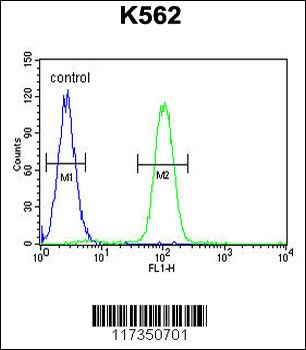
SP1 Antibody (C-term P692) flow cytometric analysis of K562 cells (right histogram) compared to a negative control cell (left histogram). FITC-conjugated goat-anti-rabbit secondary antibodies were used for the analysis.
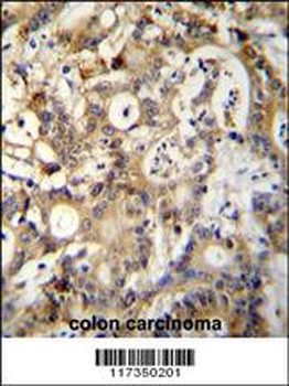
SP1 Antibody (C-term P692) immunohistochemistry analysis in formalin fixed and paraffin embedded human colon carcinoma followed by peroxidase conjugation of the secondary antibody and DAB staining. This data demonstrates the use of SP1 Antibody (C-term P692) for immunohistochemistry. Clinical relevance has not been evaluated.
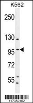
SP1 Antibody (C-term P692) western blot analysis in K562 cell line lysates (35 ug/lane). This demonstrates the SP1 antibody detected the SP1 protein (arrow).

SP1 Antibody (C-term P692) [orb1168052]
FC, IF, IHC-P, WB
Human
Rabbit
Polyclonal
Unconjugated
100 μl, 30 μlSP1 (C-term P692) Antibody [orb2630457]
FC, ICC, IHC, WB
Human, Mouse, Rat
Rabbit
Polyclonal
Unconjugated
100 μl
