You have no items in your shopping cart.
Sepharose Protein A/G
SKU: orb348756
Description
Images & Validation
−Item 1 of 3
| Tested Applications | IP, WB |
|---|---|
| Application Notes |
Key Properties
−| Purity | The product can be used for binding IgG from human, rabbit, goat, mouse and rat. It has strong binding of human, rabbit, cow, sheep, and goat polyclonal antibodies, mouse IgG2a, IgG2b, IgG3, and rat IgG2a. Protein A/G also has moderate affinity for mouse and rat IgG1 and IgG2c. It binds with weak affinity to rat IgG2b. Sepharose Protein A/G can be used for immunoprecipitation and purification of monoclonal antibodies. |
|---|---|
| Conjugation | Sepharose |
Storage & Handling
−| Storage | Store SEPHAROSE PROTEIN A/G at 4° C prior to opening. DO NOT FREEZE. |
|---|---|
| Form/Appearance | Suspension of agarose beads |
| Buffer/Preservatives | Buffer: 20% (v/v) Ethanol |
| Concentration | 0.5cc drained Sepharose per 2ml slurry |
| Hazard Information | Non-Toxic |
| Disclaimer | For research use only |
Alternative Names
−Sepharose Protein A/G, Protein AG, agarose, ProA-ProG, Staphylococcus A protein, Sepharose Protein A, Streptococcus G protein, Sepharose Protein G, immunoprecipitation, IP western blot
Quality Guarantee
Explore bioreagents carefree to elevate your research. All our products are rigorously tested for performance. If a product does not perform as described on its datasheet, our scientific support team will provide expert troubleshooting, a prompt replacement, or a refund. For full details, please see our Terms & Conditions and Buying Guide. Contact us at [email protected].
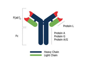
Antibody-binding protein specificity for Protein A, Protein G, and Protein A/G which demonstrate binding specificity to IgG immunoglobulin through an interaction in the Fc region of the heavy chain domain. Protein A and G each show some preferences to certain types of antibody subclasses, and proper reagent selection may be required, Protein A/G offers the widest range of antibody subclass binding. Another immunoglobulin binding protein is Protein L which binds to the kappa light chain of the F(ab')2 region.
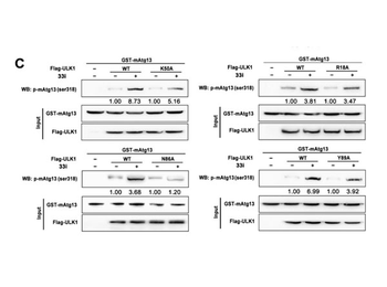
Identification of the key amino acids between ULK1 and 33i. (C) Flag-tagged ULK1WT and ULK1 mutants were expressed in HEK-293T cells and immunoprecipitated by anti-Flag antibody, then incubated with GST-tagged mAtg13 in a kinase reaction buffer in the presence or absence of 33i. The reaction was stopped and analyzed by Western blot with p-mAtg13 antibody.
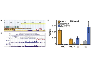
Selection and testing of species-specific PCR primers. (A) Drosophila melanogaster genome browser screen shot showing publicly available data for H3K4me2 ChIP-Seq at the eRF3 locus. (B) UCSC genome browser track for H3K4me2 ChIP-Seq in murine bone marrow derived macrophages after 3 h 100 ng/mL LPS (purple, lower track) or 16 h 1 µm dexamethasone and 3 h 100 ng/mL LPS treatment (L+D, blue, upper track). (C) ChIP-qPCR against H3K4me2 in either pure S2 cells (indicated by the fly), 25% S2 cells mixed with 75% murine macrophages treated with 100 ng/mL LPS for 3 h (marked by the fly + mouse symbol) or pure murine macrophages treated with LPS (marked by the mouse symbol). The mean of two biological replicates is plotted. Dots represent single data points, and error bars reflect the standard deviation. The color indicates the locus. (A+B) The red lines indicate the fragments amplified by PCR in C. The DNA sequence of the regions covered by the H3K4me2 signal in both species was used as input for Primer-BLAST, in order to design the primers for C.
Quick Database Links
UniProt
UniProt Details
− No UniProt data available
Documents Download
Datasheet
Product Information
Request a Document
Protocol Information
WB
Western Blot (IB, immunoblot)
IP
Immunoprecipitation
Sepharose Protein A/G (orb348756)

- 0.0
Based on 0 reviews
Participating in our Biorbyt product reviews program enables you to support fellow scientists by sharing your firsthand experience with our products.
Login to Submit a ReviewAvailable Sizes
Select a size below