You have no items in your shopping cart.
Cart summary
Item 1 of 6
Item 1 of 6
Rabbit anti-53BP1 Recombinant Monoclonal Antibody
Catalog Number: orb1519978

Product Properties
| Catalog Number | orb1519978 |
|---|---|
| Category | Antibodies |
| Description | 53BP1 Antibody |
| Target | 53BP1 |
| Clonality | Recombinant |
| Species/Host | Rabbit |
| Conjugation | Unconjugated |
| Reactivity | Human, Mouse |
| Form/Appearance | Whole IgG |
| Concentration | 100 µg/ml |
| Buffer/Preservatives | Borate Buffered Saline (BBS) pH 8.2 with 0.1% rAlbumin and 0.09% Sodium Azide |
| Immunogen | Between 350 and 400 |
| UniProt ID | Q12888 |
| Tested applications | FC, IHC, IP, WB |
| Dilution range | WB - 1:1000; IP - 20 µl/mg lysate; IHC - 1:100 to 1:500. Epitope retrieval with citrate buffer pH6.0 is recommended for FFPE tissue sections. |
| Application notes | Format: Whole IgG |
| Antibody Type | Primary Antibody |
| Clone Number | BL-250-1H11 |
| Storage | 2 - 8°C |
| Alternative names | p53-binding protein 1; tumor suppressor p53-bindin Read more... |
| Note | For research use only |
| NCBI | NP_005648.1 |
| Expiration Date | 12 months from date of receipt. |
Images

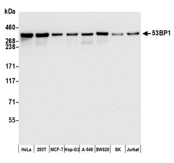
Detection of human 53BP1 by western blot. Samples: Whole cell lysate (15 µg) from HeLa, HEK293T, MCF-7, Hep-G2, A-549, SW620, SK-MEL-28 (SK) , and Jurkat cells prepared using NETN lysis buffer. Antibody: Rabbit anti-53BP1 recombinant monoclonal antibody [BL-250-1H11] (orb1519978) used at 1:1000. Secondary: HRP-conjugated goat anti-rabbit IgG. Detection: Chemiluminescence with an exposure time of 10 second.
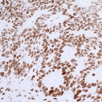
Detection of human 53BP1 by immunohistochemistry. Sample: FFPE section of colon carcinoma. Antibody: Rabbit anti-53BP1 recombinant monoclonal antibody [BL-250-1H11] (orb1519978) used at 1:250. Secondary: HRP-conjugated goat anti-rabbit IgG. Substrate: DAB.
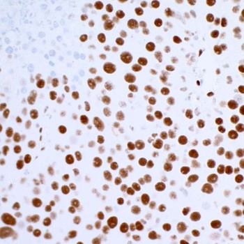
Detection of mouse 53BP1 by immunohistochemistry. Sample: FFPE section of mouse renal cell carcinoma. Antibody: Rabbit anti-53BP1 recombinant monoclonal antibody [BL-250-1H11] (orb1519978) used at 1:250. Secondary: HRP-conjugated goat anti-rabbit IgG. Substrate: DAB.
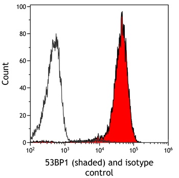
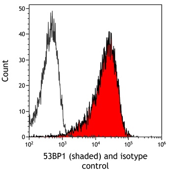
Detection of human 53BP1 (shaded) in HDLM-2 cells by flow cytometry. Antibody: Rabbit anti-53BP1 recombinant monoclonal antibody [BL-250-1H11] (orb1519978) or isotype control (unshaded). Secondary: DyLight® 650-conjugated goat anti-rabbit IgG.

Detection of mouse 53BP1 (shaded) in mIMCD-3 cells by flow cytometry. Antibody: Rabbit anti-53BP1 recombinant monoclonal antibody [BL-250-1H11] (orb1519978) or isotype control (unshaded). Secondary: DyLight® 650-conjugated goat anti-rabbit IgG.

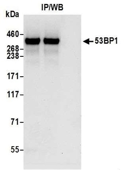
Detection of human 53BP1 by western blot of immunoprecipitates. Samples: Whole cell lysate (1.0 mg per IP reaction; 20% of IP loaded) from HEK293T cells prepared using NETN lysis buffer. Antibodies: Rabbit anti-53BP1 recombinant monoclonal antibody [BL-250-1H11] (orb1519978) used for IP at 20 µl per reaction.
Similar Products
- Item 1 of 6
Rabbit anti-53BP1 Recombinant Monoclonal Antibody [orb1519976]
FC, IHC, IP, WB
Human, Mouse
Rabbit
Recombinant
Unconjugated
20 μl 
Rabbit anti-53BP1 Recombinant Monoclonal Antibody [orb1519977]
IHC, IP, WB
Human, Mouse
Rabbit
Recombinant
Unconjugated
100 μg


Reviews
Rabbit anti-53BP1 Recombinant Monoclonal Antibody (orb1519978)

- 0.0
Based on 0 reviews
Participating in our Biorbyt product reviews program enables you to support fellow scientists by sharing your firsthand experience with our products.
Login to Submit a Review