You have no items in your shopping cart.
Cart summary
Item 1 of 3
Item 1 of 3
Profilin-1 Antibody
Catalog Number: orb1925743

Product Properties
| Catalog Number | orb1925743 |
|---|---|
| Category | Antibodies |
| Description | Profilin-1 Antibody |
| Clonality | Polyclonal |
| Species/Host | Rabbit |
| Isotype | Rabbit IgG |
| Conjugation | Unconjugated |
| Reactivity | Human, Mouse, Rat |
| Predicted Reactivity | Bovine |
| Form/Appearance | Purified polyclonal antibody supplied in PBS with 0.09% (W/V) sodium azide. This antibody is purified through a protein A column, followed by peptide affinity purification. |
| UniProt ID | P07737 |
| MW | 15054 Da |
| Tested applications | FC, WB |
| Dilution range | FC - 1:25, WB - 1:2000 |
| Storage | Maintain refrigerated at 2-8°C for up to 2 weeks. For long term storage store at -20°C in small aliquots to prevent freeze-thaw cycles |
| Research Area | Cancer Biology, Cell Biology, Neuroscience, Signal Read more... |
| Note | For research use only |
| Expiration Date | 12 months from date of receipt. |
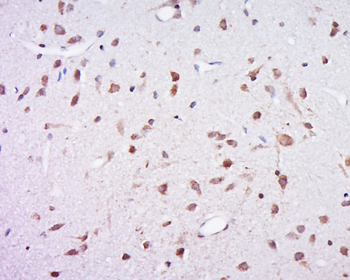
Images
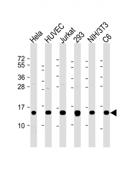
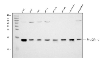
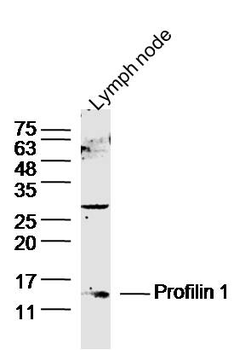
All lanes: Anti-Profilin-1 Antibody at 1:2000 dilution. Lane 1: Hela whole cell lysate. Lane 2: HUVEC whole cell lysate. Lane 3: Jurkat whole cell lysate. Lane 4: 293 whole cell lysate. Lane 5: NIH/3T3 whole cell lysate. Lane 6: C6 whole cell lysate. Lysates/proteins at 20 µg per lane. Secondary Goat Anti-Rabbit IgG, (H+L), Peroxidase conjugated at 1/10000 dilution. Predicted band size: 15 kDa. Blocking/Dilution buffer: 5% NFDM/TBST.
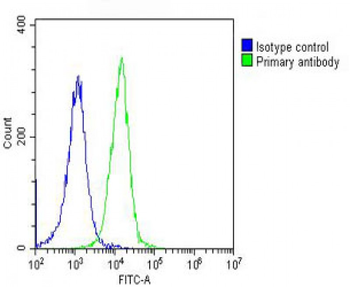
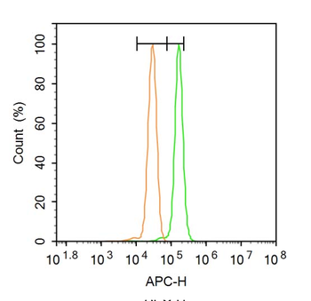
Overlay histogram showing Hela cells stained (green line). The cells were fixed with 2% paraformaldehyde (10 min) and then permeabilized with 90% methanol for 10 min. The cells were then icubated in 2% bovine serum albumin to block non-specific protein-protein interactions followed by the antibody (1:25 dilution) for 60 min at 37°C. The secondary antibody used was Goat-Anti-Rabbit IgG, DyLight 488 Conjugated Highly Cross-Adsorbed at 1/200 dilution for 40 min at 37°C. Isotype control antibody (blue line) was rabbit IgG (1 μg/1x10^6 cells) used under the same conditions. Acquisition of > 10000 events was performed.
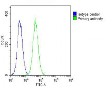
Overlay histogram showing NIH/3T3 cells stained (green line). The cells were fixed with 2% paraformaldehyde (10 min) and then permeabilized with 90% methanol for 10 min. The cells were then icubated in 2% bovine serum albumin to block non-specific protein-protein interactions followed by the antibody (1:25 dilution) for 60 min at 37°C. The secondary antibody used was Goat-Anti-Rabbit IgG, DyLight 488 Conjugated Highly Cross-Adsorbed at 1/200 dilution for 40 min at 37°C. Isotype control antibody (blue line) was rabbit IgG (1 μg/1x10^6 cells) used under the same conditions. Acquisition of > 10000 events was performed.
Similar Products
- Item 1 of 5
Profilin 1/PFN1 Antibody [orb234359]
FC, ICC, IF, IHC, WB
Human, Mouse, Rat
Rabbit
Polyclonal
Unconjugated
100 μg - Item 1 of 1
- Item 1 of 1
- Item 1 of 4
Profilin 1 Rabbit Polyclonal Antibody [orb11300]
FC, IF, IHC-Fr, IHC-P, WB
Bovine, Canine, Equine
Human, Mouse, Rat
Rabbit
Polyclonal
Unconjugated
50 μl, 200 μl, 100 μl 
Profilin 1 Rabbit Polyclonal Antibody (FITC) [orb16252]
FC, IF
Bovine, Canine, Equine
Human, Mouse, Rat
Rabbit
Polyclonal
FITC
100 μl

Reviews
Profilin-1 Antibody (orb1925743)

- 0.0
Based on 0 reviews
Participating in our Biorbyt product reviews program enables you to support fellow scientists by sharing your firsthand experience with our products.
Login to Submit a Review