You have no items in your shopping cart.
Cart summary
Item 1 of 7
Item 1 of 7
PRODH Antibody (Center)
Catalog Number: orb1928392

Product Properties
| Catalog Number | orb1928392 |
|---|---|
| Category | Antibodies |
| Description | PRODH Antibody (Center) |
| Clonality | Polyclonal |
| Species/Host | Rabbit |
| Isotype | Rabbit IgG |
| Conjugation | Unconjugated |
| Reactivity | Human, Mouse |
| Form/Appearance | Purified polyclonal antibody supplied in PBS with 0.09% (W/V) sodium azide. This antibody is purified through a protein A column, followed by peptide affinity purification. |
| UniProt ID | Q9WU79 |
| MW | 68036 Da |
| Tested applications | FC, IHC-P, WB |
| Dilution range | IHC-P - 1:100-500, WB - 1:1000 |
| Antibody Type | Primary Antibody |
| Storage | Maintain refrigerated at 2-8°C for up to 2 weeks. For long term storage store at -20°C in small aliquots to prevent freeze-thaw cycles |
| Alternative names | Pro1 |
| Research Area | Cancer Biology, Cell Biology, Signal Transduction |
| Note | For research use only |
| Expiration Date | 12 months from date of receipt. |
Images
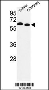
Western blot analysis of mouse Prodh Antibody (Center) in mouse liver, kidney tissue lysates (35 ug/lane). PRODH (arrow) was detected using the purified Pab.
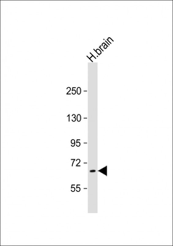
Anti-PRODH Antibody (Center) at 1:2000 dilution + human brain lysate. Lysates/proteins at 20 µg per lane. Secondary Goat Anti-Rabbit IgG, (H+L), Peroxidase conjugated at 1/10000 dilution. Predicted band size: 68 kDa. Blocking/Dilution buffer: 5% NFDM/TBST.
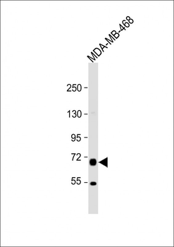
Anti-PRODH Antibody (Center) at 1:2000 dilution + MDA-MB-468 whole cell lysate. Lysates/proteins at 20 µg per lane. Secondary Goat Anti-Rabbit IgG, (H+L), Peroxidase conjugated at 1/10000 dilution. Predicted band size: 68 kDa. Blocking/Dilution buffer: 5% NFDM/TBST.
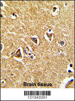
Formalin-fixed and paraffin-embedded human brain tissue reacted with mouse Prodh Antibody (Center), which was peroxidase-conjugated to the secondary antibody, followed by DAB staining. This data demonstrates the use of this antibody for immunohistochemistry; clinical relevance has not been evaluated.
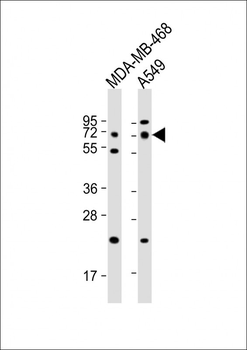
All lanes: Anti-PRODH Antibody (Center) at 1:1000 dilution. Lane 1: MDA-MB-468 whole cell lysates. Lane 2: A549 whole cell lysates. Lysates/proteins at 20 µg per lane. Secondary Goat Anti-Rabbit IgG, (H+L), Peroxidase conjugated at 1/10000 dilution. Predicted band size: 68 kDa. Blocking/Dilution buffer: 5% NFDM/TBST.

Staining PRODH in H. skeletal muscle sections by Immunohistochemistry (IHC-P - paraformaldehyde-fixed, paraffin-embedded sections). Tissue was fixed with formaldehyde and blocked with 3% BSA for 0.5 hour at room temperature; antigen retrieval was by heat mediation with a citrate buffer (pH6). Samples were incubated with primary antibody (1/25) for 1 hours at 37°C. A undiluted biotinylated goat polyvalent antibody was used as the secondary antibody.
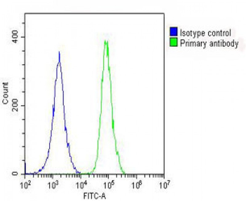
Overlay histogram showing A549 cells stained (green line). The cells were fixed with 2% paraformaldehyde (10 min) and then permeabilized with 90% methanol for 10 min. The cells were then icubated in 2% bovine serum albumin to block non-specific protein-protein interactions followed by the antibody (1:25 dilution) for 60 min at 37°C. The secondary antibody used was Goat-Anti-Rabbit IgG, DyLight 488 Conjugated Highly Cross-Adsorbed at 1/200 dilution for 40 min at 37°C. Isotype control antibody (blue line) was rabbit IgG (1 μg/1x10^6 cells) used under the same conditions. Acquisition of > 10000 events was performed.
Reviews
PRODH Antibody (Center) (orb1928392)

- 0.0
Based on 0 reviews
Participating in our Biorbyt product reviews program enables you to support fellow scientists by sharing your firsthand experience with our products.
Login to Submit a Review