You have no items in your shopping cart.
Cart summary
Item 1 of 2
Item 1 of 2
p53 Antibody / TP53 (N-Terminal Region)
Catalog Number: orb749391

Product Properties
| Catalog Number | orb749391 |
|---|---|
| Category | Antibodies |
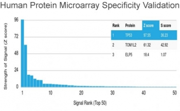
| Description | This antibody is specific for a 53kDa protein, which is identified as p53 suppressor gene product. It reacts with the mutant as well as the wild form of p53 under denaturing and non-denaturing conditions. The antibody epitope maps within the N-terminus (aa 20-25) of p53 oncoprotein. p53 is a tumor suppressor gene expressed in a wide variety of tissue types and is involved in regulating cell growth, replication, and apoptosis. It binds to MDM2, SV40 T antigen and human papilloma virus E6 protein. Positive nuclear staining with p53 antibody has been reported to be a negative prognostic factor in breast carcinoma, lung carcinoma, colorectal, and urothelial carcinoma. antibody to p53 positivity has also been used to differentiate uterine serous carcinoma from endometrioid carcinoma as well as to detect intratubular germ cell neoplasia. Mutations involving p53 are found in a wide variety of malignant tumors, including breast, ovarian, bladder, colon, lung, and melanoma. |
| Clonality | Monoclonal |
| Species/Host | Mouse |
| Isotype | Mouse IgG2a, kappa |
| Conjugation | Unconjugated |
| Reactivity | Human |
| Immunogen | Recombinant human wild-type p53 protein was used as the immunogen for this antibody. |
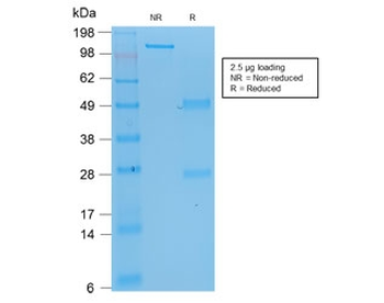
| Tested applications | IHC-P, WB |
| Dilution range | Western blot: 1-2ug/ml,Immunohistochemistry (FFPE): 1-2ug/ml for 30 min at RT |
| Application notes | The concentration stated for each application is a general starting point. Variations in protocols, secondaries and substrates may require the antibody to be titered up or down for optimal performance.1. Staining of formalin-fixed tissues requires boiling tissue sections in pH 9 10mM Tris with 1mM EDTA for 10-20 min followed by cooling at RT for 20 minutes.2. The prediluted format is supplied in a dropper bottle and is optimized for use in IHC. After epitope retrieval step (if required), drip mAb solution onto the tissue section and incubate at RT for 30 min. |
| Antibody Type | Primary Antibody |
| Clone Number | BP53-12 |
| Formula | 0.2 mg/ml in 1X PBS with 0.1 mg/ml BSA (US sourced) and 0.05% sodium azide |
| Storage | Maintain refrigerated at 2-8°C for up to 2 weeks. For long term storage store at -20°C in small aliquots to prevent freeze-thaw cycles. |
| Note | For research use only |
| Expiration Date | 12 months from date of receipt. |
Images
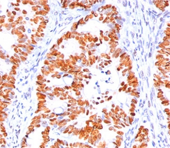
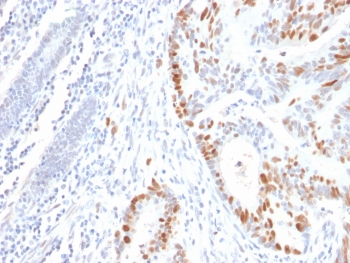
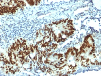
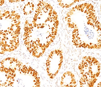
IHC staining of human colon carcinoma with p53 antibody (clone BP53-12).
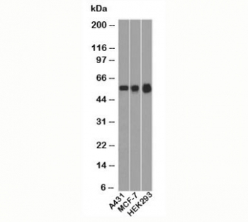
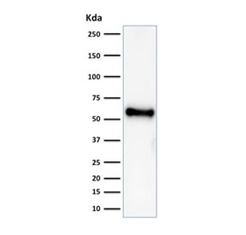
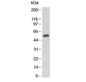
Western blot testing of human samples with p53 antibody (clone BP53-12).
Similar Products
- Item 1 of 4
p53 Antibody / TP53 N-Terminal Region [orb606470]
IHC-P, WB
Human
Mouse
Recombinant
Unconjugated
20 μg, 100 μg - Item 1 of 3
p53 Antibody / TP53 (N-Terminal Region) [orb402962]
FACS, IF, IHC-P, WB
Human
Mouse
Monoclonal
Unconjugated
20 μg, 100 μg - Item 1 of 2
p53 Antibody / TP53 (N-Terminal Region) [orb749392]
IHC-P, WB
Human
Mouse
Monoclonal
Unconjugated
20 μg, 100 μg

Reviews
p53 Antibody / TP53 (N-Terminal Region) (orb749391)

- 0.0
Based on 0 reviews
Participating in our Biorbyt product reviews program enables you to support fellow scientists by sharing your firsthand experience with our products.
Login to Submit a Review