You have no items in your shopping cart.
Cart summary

| Catalog Number | orb769198 |
|---|---|
| Category | Antibodies |
| Description | Rabbit polyclonal antibody to MYPT1 (phospho-Thr853) |
| Clonality | Polyclonal |
| Isotype | IgG |
| Conjugation | Unconjugated |
| Reactivity | Human, Mouse, Rat |
| Concentration | 1 mg/ml |
| Buffer/Preservatives | PBS with 0.02% sodium azide and 50% glycerol pH 7.4. |
| Purification | The antibody was affinity-purified from rabbit antiserum by affinity-chromatography using epitope-specific immunogen. |
| Immunogen | The antiserum was produced against synthesized peptide derived from human MYPT1 around the phosphorylation site of Thr853. AA range:621-670 |
| MW | 130 |
| Tested applications | ELISA, IF, IHC-P, WB |
| Dilution range | IF: 1:50-200 Western Blot: 1/500 - 1/2000. Immunohistochemistry: 1/100 - 1/300. ELISA: 1/5000. Not yet tested in other applications. |
| Source | Rabbit |
| Storage | Maintain refrigerated at 2-8°C for up to 2 weeks. For long term storage store at -20°C in small aliquots to prevent freeze-thaw cycles. |
| Alternative names | Anti-PPP1R12A antibody, anti-MBS antibody, anti-MY Read more... |
| Note | For research use only |
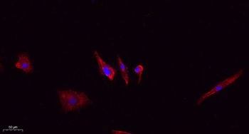
Immunofluorescence analysis of A549. 1, primary Antibody (red) was diluted at 1:200 (4°C overnight). 2, Goat Anti Rabbit IgG (H&L) - Alexa Fluor 594 Secondary antibody was diluted at 1:1000 (room temperature, 50min). 3, Picture B: DAPI (blue) 10min.
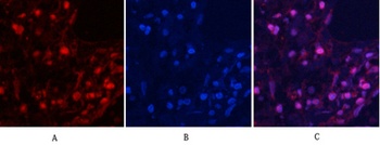
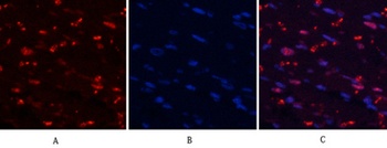
Immunofluorescence analysis of human-lung tissue. 1, MYPT1 (phospho Thr853) Polyclonal Antibody (red) was diluted at 1:200 (4°C, overnight). 2, Cy3 labled Secondary antibody was diluted at 1:300 (room temperature, 50min). 3, Picture B: DAPI (blue) 10min. Picture A:Target. Picture B: DAPI. Picture C: merge of A + B.
Immunofluorescence analysis of human-lung tissue. 1, MYPT1 (phospho Thr853) Polyclonal Antibody (red) was diluted at 1:200 (4°C, overnight). 2, Cy3 labled Secondary antibody was diluted at 1:300 (room temperature, 50min). 3, Picture B: DAPI (blue) 10min. Picture A:Target. Picture B: DAPI. Picture C: merge of A + B.
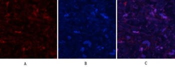
Immunofluorescence analysis of rat-heart tissue. 1, MYPT1 (phospho Thr853) Polyclonal Antibody (red) was diluted at 1:200 (4°C, overnight). 2, Cy3 labled Secondary antibody was diluted at 1:300 (room temperature, 50min). 3, Picture B: DAPI (blue) 10min. Picture A:Target. Picture B: DAPI. Picture C: merge of A + B.

Phospho-MYPT1 (Thr853) Rabbit pAb, PE-Cy5 conjugated [orb2843942]
IF
Bovine, Canine, Equine, Gallus, Rabbit, Rat
Human, Mouse
Rabbit
Polyclonal
PE/Cy5
100 μlPhospho-MYPT1 (Thr853) Rabbit pAb, PE-Cy5.5 conjugated [orb2843943]
IF
Bovine, Canine, Equine, Gallus, Rabbit, Rat
Human, Mouse
Rabbit
Polyclonal
PE/Cy5.5
100 μlPhospho-MYPT1 (Thr853) Rabbit pAb, PE-Cy7 conjugated [orb2843944]
IF
Bovine, Canine, Equine, Gallus, Rabbit, Rat
Human, Mouse
Rabbit
Polyclonal
PE/Cy7
100 μlPhospho-MYPT1 (Thr853) Rabbit pAb, PerCP-Cy7 conjugated [orb2843945]
IF
Bovine, Canine, Equine, Gallus, Rabbit, Rat
Human, Mouse
Rabbit
Polyclonal
PerCP/Cy7
100 μlPhospho-MYPT1 (Thr853) Rabbit pAb, Pacific Blue conjugated [orb2843946]
IF
Bovine, Canine, Equine, Gallus, Rabbit, Rat
Human, Mouse
Rabbit
Polyclonal
Pacific Blue
100 μl
