You have no items in your shopping cart.
Cart summary
Item 1 of 2
Item 1 of 2
HSP90 Antibody
Catalog Number: orb1822502

Product Properties
| Catalog Number | orb1822502 |
|---|---|
| Category | Antibodies |
| Description | Mouse Anti-Water Mold HSP90 Monoclonal IgG2b |
| Target | HSP90 |
| Clonality | Monoclonal |
| Species/Host | Mouse |
| Isotype | IgG2b |
| Conjugation | Unconjugated |
| Reactivity | Fungi, Gallus, Human, Insect, Invertebrate, Mouse, Plant, Rabbit, Rat |
| Concentration | 1 mg/ml |
| Buffer/Preservatives | PBS pH 7.4, 50% glycerol, 0.09% sodium azide *Storage buffer changes when conjugated |
| Purification | Protein G Purified |
| Immunogen | Heat shock protein 90 from the water mold Achyla ambisexualis |
| UniProt ID | Q8LLI5 |
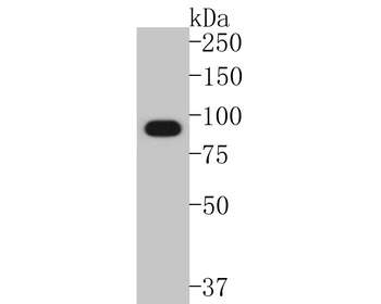
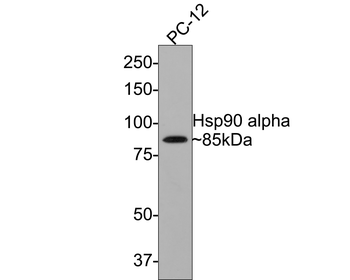
| MW | 90kDa |
| Tested applications | AM, IHC, WB |
| Dilution range | WB (1:1000), IHC (1:2000); optimal dilutions for assays should be determined by the user. |
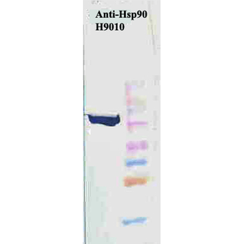
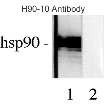
| Application notes | 1 µg/ml of SMC-112 was sufficient for detection of HSP90 in 20 µg of heat shocked HeLa cell lysate by colorimetric immunoblot analysis using Goat anti-mouse IgG:HRP as the secondary antibody |
| Clone Number | AC-16 |
| Storage | Maintain refrigerated at 2-8°C for up to 2 weeks. For long term storage store at -20°C in small aliquots to prevent freeze-thaw cycles. |
| Alternative names | HSP84 Antibody, HSP86 Antibody, HSP90 Antibody, HS Read more... |
| Research Area | Cancer Biology, Cell Biology, Protein Biochemistry Read more... |
| Note | For research use only |
| Entrez | 4768 |
| NCBI | AAM90675.1 |
Images
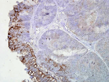
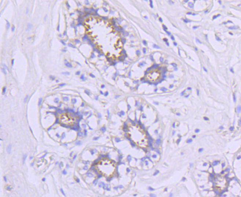
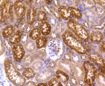
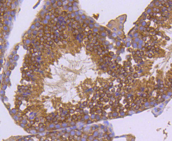
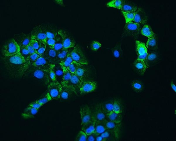
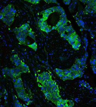
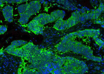
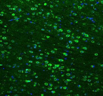
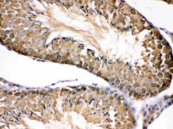
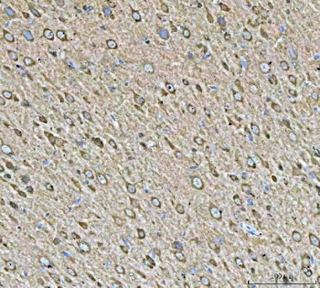
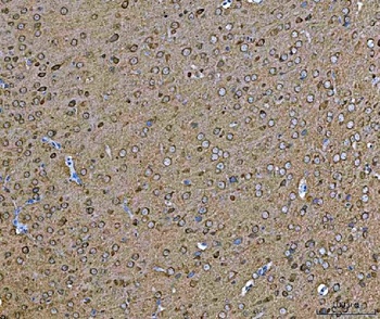
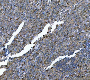
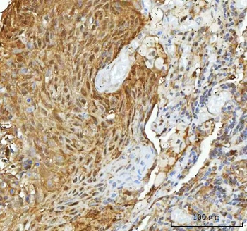
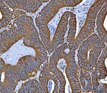
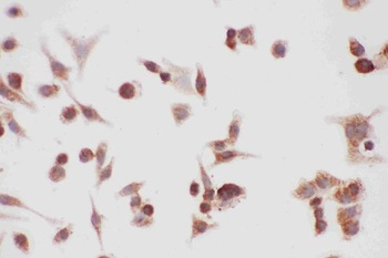
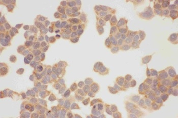
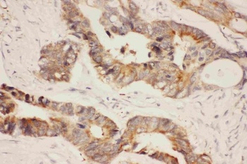
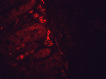
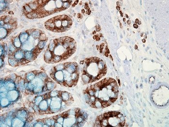
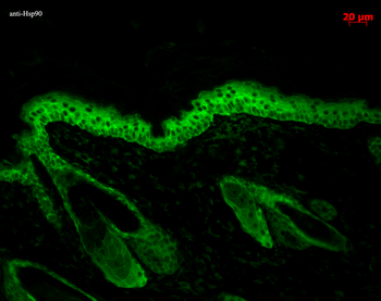
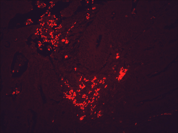
Immunohistochemistry analysis using Mouse Anti-Hsp90 Monoclonal Antibody, Clone AC-16. Tissue: colon carcinoma. Species: Human. Fixation: Formalin. Primary Antibody: Mouse Anti-Hsp90 Monoclonal Antibody at 1:2000 for 12 hours at 4°C. Secondary Antibody: Biotin Goat Anti-Mouse at 1:2000 for 1 hour at RT. Counterstain: Mayer Hematoxylin (purple/blue) nuclear stain at 200 μl for 2 minutes at RT. Localization: Inflammatory cells. Magnification: 40x. Inflammatory cells.
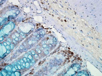
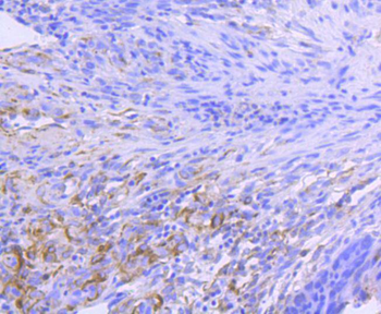
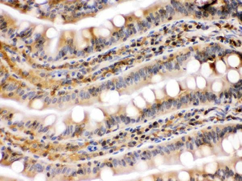
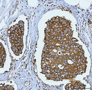
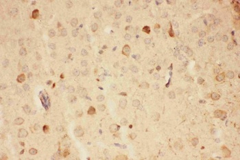
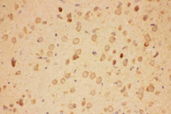
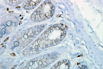
Immunohistochemistry analysis using Mouse Anti-Hsp90 Monoclonal Antibody, Clone AC-16. Tissue: inflamed colon. Species: Mouse. Fixation: Formalin. Primary Antibody: Mouse Anti-Hsp90 Monoclonal Antibody at 1:2000 for 12 hours at 4°C. Secondary Antibody: Biotin Goat Anti-Mouse at 1:2000 for 1 hour at RT. Counterstain: Mayer Hematoxylin (purple/blue) nuclear stain at 200 μl for 2 minutes at RT. Localization: Inflammatory cells. Magnification: 40x. Mostly inflammatory cells, some mucosa.
Similar Products
- Item 1 of 10
Hsp90 alpha Recombinant Rabbit Monoclonal Antibody [orb1499397]
ICC, IF, IHC-Fr, IHC-P, WB
Human, Mouse, Rat
Human, Mouse, Rat
Rabbit
Recombinant
Unconjugated
25 μl, 100 μl, 50 μl - Item 1 of 9
Hsp90 beta/HSP90AB1 Antibody [orb315144]
ICC, IF, IHC, WB
Human, Mouse, Rat
Rabbit
Polyclonal
Unconjugated
100 μg - Item 1 of 8
Hsp90 alpha + beta HSP90AA1 Rabbit Monoclonal Antibody [orb547614]
FC, ICC, IF, IHC, IP, WB
Human, Mouse, Rat
Rabbit
Monoclonal
Unconjugated
100 μl - Item 1 of 8
Hsp90 alpha/HSP90AA1 Antibody [orb196259]
FC, ICC, IF, IHC, WB
Human, Mouse, Rat
Rabbit
Polyclonal
Unconjugated
100 μg - Item 1 of 8
HSP90 Antibody: APC [orb146802]
ICC, IF, IHC
Canine, Fish, Gallus, Hamster, Human, Mouse, Rabbit, Rat
Mouse
Monoclonal
APC
200 μg

Reviews
HSP90 Antibody (orb1822502)

- 0.0
Based on 0 reviews
Participating in our Biorbyt product reviews program enables you to support fellow scientists by sharing your firsthand experience with our products.
Login to Submit a Review