You have no items in your shopping cart.
Cart summary
Item 1 of 7
Item 1 of 7
Goat anti-IFNGR1 (aa181-193) Antibody
Catalog Number: orb107681

Product Properties
| Catalog Number | orb107681 |
|---|---|
| Category | Antibodies |
| Description | Goat polyclonal to IFNGR1 |
| Target | IFNGR1 (aa181-193) |
| Clonality | Polyclonal |
| Species/Host | Goat |
| Conjugation | Unconjugated |
| Reactivity | Human |
| Buffer/Preservatives | Supplied at 0.5 mg/ml in Tris saline, 0.02% sodium azide, pH 7.3 with 0.5% bovine serum albumin. Aliquot and store at -20°C. Minimize freezing and thawing. |
| Purification | Purified from goat serum by ammonium sulphate precipitation followed by antigen affinity chromatography using the immunizing peptide. |
| Protein Sequence | SEIQYKILTQKED |
| MW | 54.4 |
| Tested applications | ELISA, FC, IF, IHC, WB |
| Dilution range | ELISA: 1:128000, WB: 0.3-1 μg/ml |
| Application notes | WB: Approx 70kDa band observed in lysates of cell line HepG2 (calculated MW of 54.4kDa according to NP_000407.1). The observed molecular weight corresponds to the glycosylated form. Recommended concentration: 0.3-1µg/ml. |
| Storage | Maintain refrigerated at 2-8°C for up to 2 weeks. For long term storage store at -20°C in small aliquots to prevent freeze-thaw cycles. |
| Alternative names | anti IFNGR1 antibody |
| Research Area | Stem Cells |
| Note | For research use only |
| Entrez | 3459 |
Images
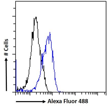
Flow cytometric analysis of paraformaldehyde fixed K562 cells (blue line), permeabilized with 0.5% Triton. Primary incubation 1hr (10 ug/ml) followed by Alexa Fluor 488 secondary antibody (1 ug/ml). IgG control: Unimmunized goat IgG (black line) followed by Alexa Fluor 488 secondary antibody.
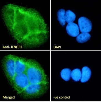
Immunofluorescence analysis of paraformaldehyde fixed Caco-2 cells, permeabilized with 0.15% Triton. Primary incubation 1hr (10 ug/ml) followed by Alexa Fluor 488 secondary antibody (2 ug/ml), showing membrane staining. The nuclear stain is DAPI (blue). Negative control: Unimmunized goat IgG (10 ug/ml) followed by Alexa Fluor 488 secondary antibody (2 ug/ml).
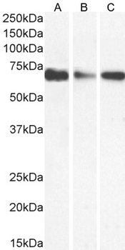
1 µg/ml staining of K562 (A) Caco-2 (B) and HepG2 (C) cell lysate (35 µg protein in RIPA buffer). Detected by chemiluminescence.
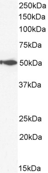
2 µg/ml staining of Human Spleen lysate (35 µg protein in RIPA buffer). Detected by chemiluminescence.
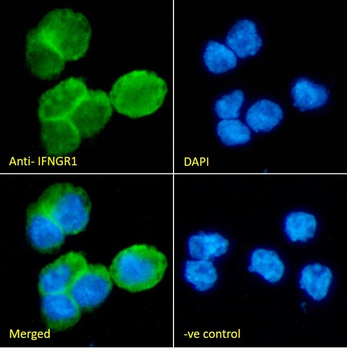
Immunofluorescence analysis of paraformaldehyde fixed THP-1 cells immobilized on ShifixTM coverslip, permeabilized with 0.15% Triton. Primary incubation 1hr (10 ug/ml) followed by Alexa Fluor 488 secondary antibody (2 ug/ml), showing membrane and cytoplasmic staining. The nuclear stain is DAPI (blue). Negative control: Unimmunized goat IgG (10 ug/ml) followed by Alexa Fluor 488 secondary antibody (2 ug/ml).
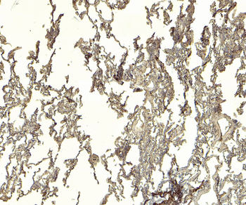
4 µg/ml staining of paraffin embedded Human Lung. Heat induced antigen retrieval with citrate buffer pH6, HRP-staining.
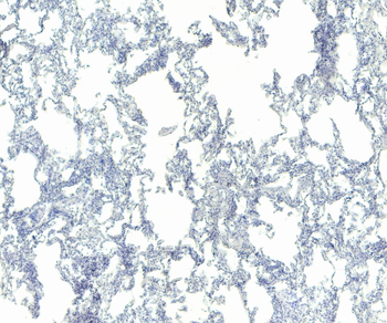
Negative Control showing staining of paraffin embedded Human Lung, with no primary antibody.
Reviews
Goat anti-IFNGR1 (aa181-193) Antibody (orb107681)

- 0.0
Based on 0 reviews
Participating in our Biorbyt product reviews program enables you to support fellow scientists by sharing your firsthand experience with our products.
Login to Submit a Review