You have no items in your shopping cart.
Cart summary
Item 1 of 6
Item 1 of 6
Furin Recombinant Rabbit Monoclonal Antibody
Catalog Number: orb1499355

Product Properties
| Catalog Number | orb1499355 |
|---|---|
| Category | Antibodies |
| Description | Furin Recombinant Rabbit Monoclonal Antibody |
| Target | FURIN |
| Clonality | Recombinant |
| Species/Host | Rabbit |
| Isotype | IgG |
| Conjugation | Unconjugated |
| Reactivity | Human, Mouse |
| Predicted Reactivity | Rat |
| Form/Appearance | Liquid |
| Concentration | 1mg/ml |
| Buffer/Preservatives | 0.01M TBS (pH7.4) with 1% rAlbumin, 0.02% Proclin300 and 50% Glycerol. |
| Purification | Affinity purified by Protein A |
| Immunogen | KLH conjugated synthetic peptide derived from human Furin |
| UniProt ID | P09958 |
| MW | 74 kDa |
| Tested applications | IF, IHC-Fr, IHC-P, WB |
| Dilution range | WB=1:500-200, IHC-P=1:100-500, IHC-F=1:400-800, IF=1:100-500 |
| Antibody Type | Primary Antibody |
| Storage | Maintain refrigerated at 2-8°C for up to 2 weeks. For long term storage store at -20°C in small aliquots to prevent freeze-thaw cycles. |
| Alternative names | FURIN_HUMAN; Furin; EC:3.4.21.75; FURIN; FUR; PACE Read more... |
| Note | For research use only |
| Expiration Date | 12 months from date of receipt. |
Images
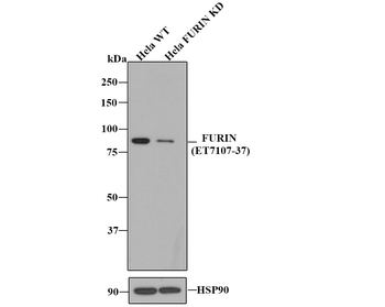
All lanes: Western blot analysis of FURIN with anti-Furin antibody (orb1499355) at 1:500 dilution. Lane 1: Wild-type Hela whole cell lysate. Lane 2: FURIN knockdown Hela whole cell lysate.
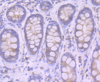
Immunohistochemical analysis of paraffin-embedded human colon tissue using anti-Furin antibody. The section was pre-treated using heat mediated antigen retrieval with Tris-EDTA buffer (pH 8.0-8.4) for 20 minutes. The tissues were blocked in 5% BSA for 30 minutes at room temperature, washed with ddH2O and PBS, and then probed with the primary antibody (orb1499355, 1/50) for 30 minutes at room temperature. The detection was performed using an HRP conjugated compact polymer system. DAB was used as the chromogen. Tissues were counterstained with hematoxylin and mounted with DPX.
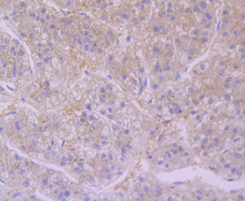
Immunohistochemical analysis of paraffin-embedded human liver tissue using anti-Furin antibody. The section was pre-treated using heat mediated antigen retrieval with Tris-EDTA buffer (pH 8.0-8.4) for 20 minutes. The tissues were blocked in 5% BSA for 30 minutes at room temperature, washed with ddH2O and PBS, and then probed with the primary antibody (orb1499355, 1/50) for 30 minutes at room temperature. The detection was performed using an HRP conjugated compact polymer system. DAB was used as the chromogen. Tissues were counterstained with hematoxylin and mounted with DPX.
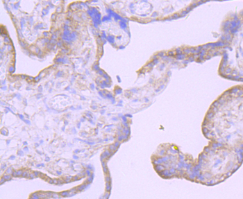
Immunohistochemical analysis of paraffin-embedded human placenta tissue using anti-Furin antibody. The section was pre-treated using heat mediated antigen retrieval with Tris-EDTA buffer (pH 8.0-8.4) for 20 minutes. The tissues were blocked in 5% BSA for 30 minutes at room temperature, washed with ddH2O and PBS, and then probed with the primary antibody (1/50) for 30 minutes at room temperature. The detection was performed using an HRP conjugated compact polymer system. DAB was used as the chromogen. Tissues were counterstained with hematoxylin and mounted with DPX.
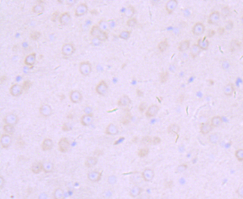
Immunohistochemical analysis of paraffin-embedded mouse brain tissue using anti-Furin antibody. The section was pre-treated using heat mediated antigen retrieval with Tris-EDTA buffer (pH 8.0-8.4) for 20 minutes. The tissues were blocked in 5% BSA for 30 minutes at room temperature, washed with ddH2O and PBS, and then probed with the primary antibody (orb1499355, 1/50) for 30 minutes at room temperature. The detection was performed using an HRP conjugated compact polymer system. DAB was used as the chromogen. Tissues were counterstained with hematoxylin and mounted with DPX.
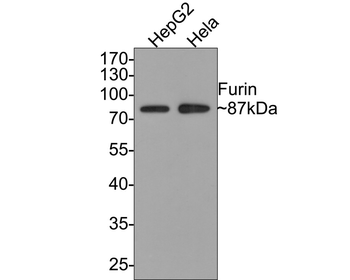
Western blot analysis of Furin on different lysates with Rabbit anti-Furin antibody (orb1499355) at 1/500 dilution. Lane 1: HepG2 cell lysate, Lane 2: Hela cell lysate, Lysates/proteins at 10 µg/Lane. Predicted band size: 87 kDa, Observed band size: 87 kDa, Exposure time: 1 minute, 10% SDS-PAGE gel.
Similar Products

Recombinant FURIN Rabbit mAb Antibody [orb2989630]
WB
Human, Rat
Rabbit
Monoclonal
Unconjugated
30 μl, 50 μl, 100 μl, 200 μl
Reviews
Furin Recombinant Rabbit Monoclonal Antibody (orb1499355)

- 0.0
Based on 0 reviews
Participating in our Biorbyt product reviews program enables you to support fellow scientists by sharing your firsthand experience with our products.
Login to Submit a Review