You have no items in your shopping cart.
Cart summary
Item 1 of 5
Item 1 of 5
DDR1 Antibody
Catalog Number: orb1263030

| Catalog Number | orb1263030 |
|---|---|
| Category | Antibodies |
| Description | DDR1 Antibody |
| Target | DDR1 |
| Clonality | Polyclonal |
| Isotype | Rabbit Ig |
| Conjugation | Unconjugated |
| Reactivity | Human, Rat |
| Predicted Reactivity | Mouse |
| Form/Appearance | Liquid |
| Concentration | batch dependent |
| Buffer/Preservatives | Supplied in PBS with 0.09% (W/V) sodium azide. |
| Purification | This antibody is purified through a protein A column, followed by peptide affinity purification. |
| Immunogen | This DDR1 antibody is generated from rabbits immunized with a KLH conjugated synthetic peptide between 17-47 amino acids from the N-terminal region of human DDR1. |
| UniProt ID | Q08345 |
| MW | 101 kDa |
| Tested applications | FC, IHC-P, WB |
| Application notes | For FACS starting dilution is: 1:25For WB starting dilution is: 1:2000For IHC-P starting dilution is: 1:10~50 |
| Antibody Type | Primary Antibody |
| Storage | Maintain refrigerated at 2-8°C for up to 2 weeks. For long term storage store at -20°C in small aliquots to prevent freeze-thaw cycles. |
| Alternative names | Epithelial discoidin domain-containing receptor 1, Read more... |
| Note | For research use only |
| NCBI | Q08345 |

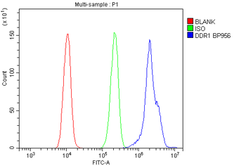
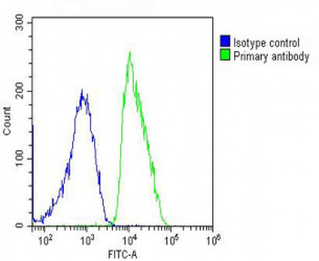
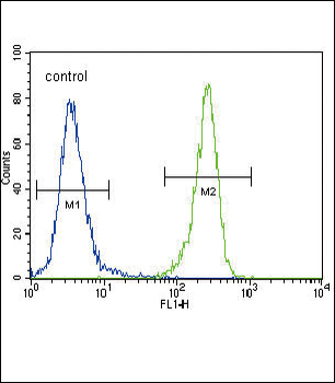
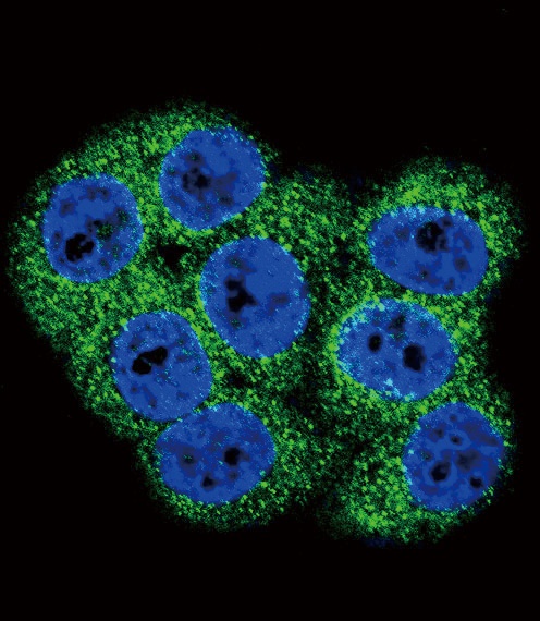
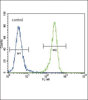
Overlay histogram showing MCF-7 cells stained with Antibody (green line). The cells were fixed with 2% paraformaldehyde (10 min) and then permeabilized with 90% methanol for 10 min. The cells were then icubated in 2% bovine serum albumin to block non-specific protein-protein interactions followed by the antibody (1:25 dilution) for 60 min at 37°C. The secondary antibody used was Goat-Anti-Rabbit IgG, Conjugated Highly Cross-Adsorbed at 1/200 dilution for 40 min at 37°C. Isotype control antibody (blue line) was rabbit IgG (1ug/1x10^6 cells) used under the same conditions. Acquisition of > 10000 events was performed.

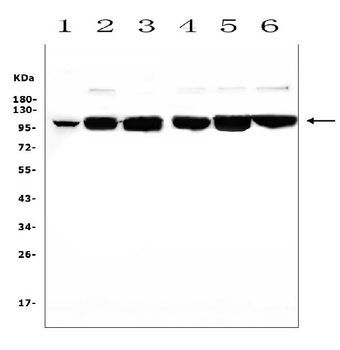
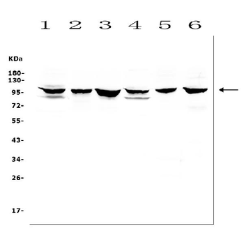
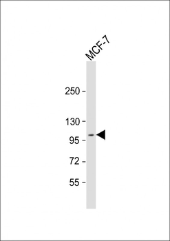
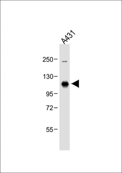
Western Blot at 1:2000 dilution + MCF-7 whole cell lysate Lysates/proteins at 20 ug per lane.
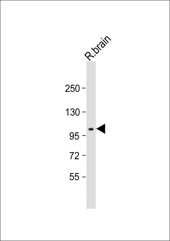
Western Blot at 1:2000 dilution + rat brain lysate Lysates/proteins at 20 ug per lane.
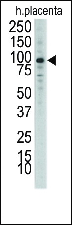
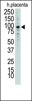
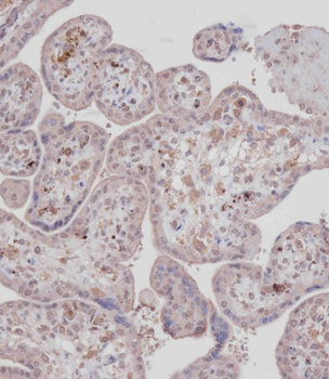
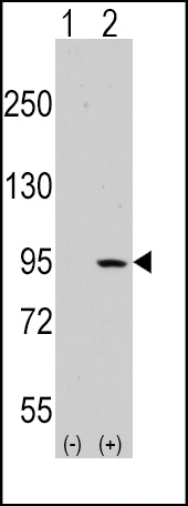
Western blot analysis of anti-DDR1 Antibody Pab in placenta lysate
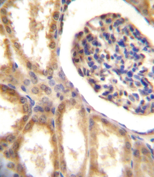
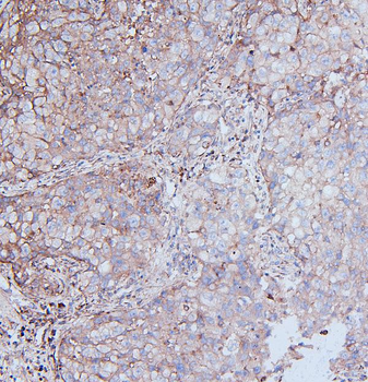
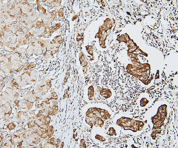
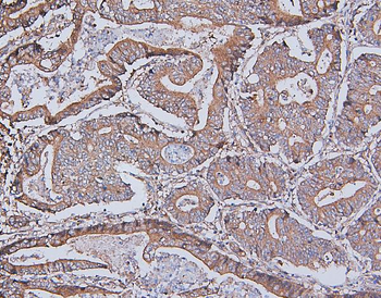
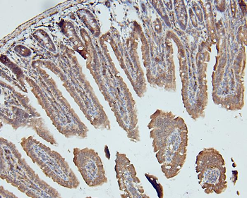
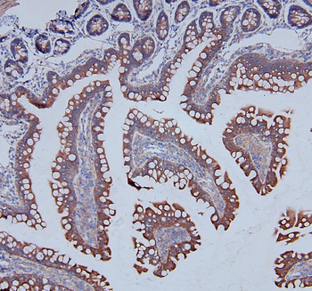
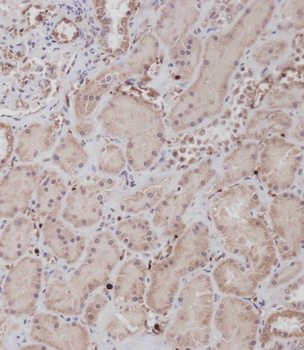
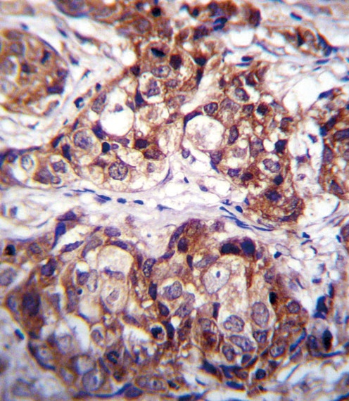
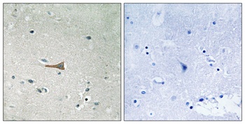
DDR1 Antibody immunohistochemistry analysis in formalin fixed and paraffin embedded human kidney tissue followed by peroxidase conjugation of the secondary antibody and DAB staining.
- Item 1 of 9
MCK10/NEP/DDR1 Antibody [orb570379]
ELISA, FC, ICC, IF, IHC, WB
Human, Mouse, Rat
Rabbit
Polyclonal
Unconjugated
10 μg, 100 μg - Item 1 of 5
DDR1 Antibody (N-term) [orb1929015]
FC, IHC-P, WB
Mouse
Human, Rat
Rabbit
Polyclonal
Unconjugated
100 μl, 50 μl - Item 1 of 5
DDR1 Antibody (Center) [orb1929386]
FC, IF, IHC-P, WB
Human
Rabbit
Polyclonal
Unconjugated
100 μl, 50 μl - Item 1 of 4
- Item 1 of 4
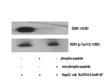
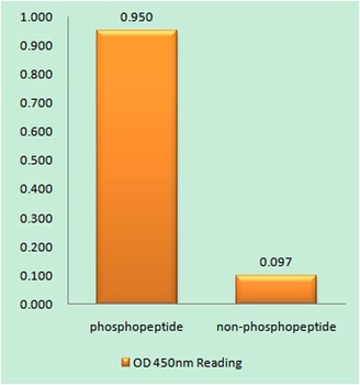
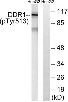
DDR1 (phospho Tyr513) rabbit pAb [orb764372]
ELISA, IHC-P, WB
Human, Mouse, Rat
Polyclonal
Unconjugated
50 μl, 100 μl