You have no items in your shopping cart.
Cart summary
Item 1 of 7
Item 1 of 7
BRD2 Recombinant Rabbit Monoclonal Antibody
Catalog Number: orb1499352

Product Properties
| Catalog Number | orb1499352 |
|---|---|
| Category | Antibodies |
| Description | BRD2 Recombinant Rabbit Monoclonal Antibody |
| Target | BRD2 |
| Clonality | Recombinant |
| Species/Host | Rabbit |
| Isotype | IgG |
| Conjugation | Unconjugated |
| Reactivity | Human |
| Predicted Reactivity | Human |
| Form/Appearance | Liquid |
| Concentration | 1mg/ml |
| Buffer/Preservatives | 0.01M TBS (pH7.4) with 1% rAlbumin, 0.02% Proclin300 and 50% Glycerol. |
| Purification | Affinity purified by Protein A |
| Immunogen | A synthesized peptide derived from human BRD2 (1-55/801aa) |
| UniProt ID | P25440 |
| MW | 88 kDa |
| Tested applications | FC, ICC, IF, IHC-Fr, IHC-P, WB |
| Dilution range | WB=1:500-2000, IHC-P=1:100-500, IHC-F=1:100-500, ICC/IF=1:50-200, IF=1:100-500, Flow-Cyt=1:50-200 |
| Antibody Type | Primary Antibody |
| Clone Number | R2A5 |
| Storage | Maintain refrigerated at 2-8°C for up to 2 weeks. For long term storage store at -20°C in small aliquots to prevent freeze-thaw cycles. |
| Alternative names | Bromodomain-containing protein 2; KIAA9001; RING3; Read more... |
| Research Area | Bromodomains (BRDs), Cell Biology, Developmental B Read more... |
| Note | For research use only |
| Expiration Date | 12 months from date of receipt. |
Images
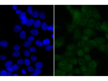
ICC staining of BRD2 in 293T cells (green). Formalin fixed cells were permeabilized with 0.1% Triton X-100 in TBS for 10 minutes at room temperature and blocked with 10% negative goat serum for 15 minutes at room temperature. Cells were probed with the primary antibody (orb1499352, 1/50) for 1 hour at room temperature, washed with PBS. Alexa Fluor®488 conjugate-Goat anti-Rabbit IgG was used as the secondary antibody at 1/1000 dilution. The nuclear counter stain is DAPI (blue).
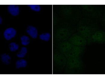
ICC staining of BRD2 in A431 cells (green). Formalin fixed cells were permeabilized with 0.1% Triton X-100 in TBS for 10 minutes at room temperature and blocked with 10% negative goat serum for 15 minutes at room temperature. Cells were probed with the primary antibody (orb1499352, 1/50) for 1 hour at room temperature, washed with PBS. Alexa Fluor®488 conjugate-Goat anti-Rabbit IgG was used as the secondary antibody at 1/1000 dilution. The nuclear counter stain is DAPI (blue).
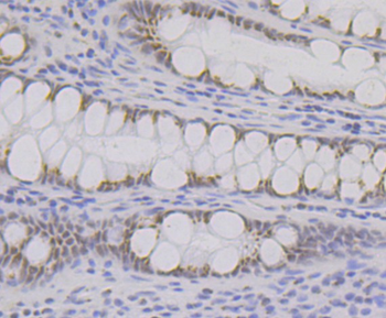
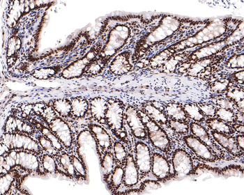
Immunohistochemical analysis of paraffin-embedded human colon tissue using anti-BRD2 antibody. The section was pre-treated using heat mediated antigen retrieval with Tris-EDTA buffer (pH9.0) for 20 minutes. The tissues were blocked in 1% BSA for 30 minutes at room temperature, washed with ddH2O and PBS, and then probed with the primary antibody (orb1499352, 1/50) for 30 minutes at room temperature. The detection was performed using an HRP conjugated compact polymer system. DAB was used as the chromogen. Tissues were counterstained with hematoxylin and mounted with DPX.
Immunohistochemical analysis of paraffin-embedded human gallbladder tissue using anti-BRD2 antibody. The section was pre-treated using heat mediated antigen retrieval with sodium citrate buffer (pH 6.0) for 20 minutes. The tissues were blocked in 1% BSA for 30 minutes at room temperature, washed with ddH2O and PBS, and then probed with the primary antibody (orb1499352, 1/100) for 30 minutes at room temperature. The detection was performed using an HRP conjugated compact polymer system. DAB was used as the chromogen. Tissues were counterstained with hematoxylin and mounted with DPX.
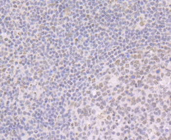
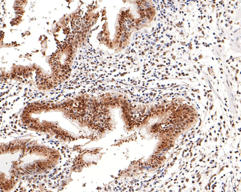
Immunohistochemical analysis of paraffin-embedded human tonsil tissue using anti-BRD2 antibody. The section was pre-treated using heat mediated antigen retrieval with Tris-EDTA buffer (pH9.0) for 20 minutes. The tissues were blocked in 1% BSA for 30 minutes at room temperature, washed with ddH2O and PBS, and then probed with the primary antibody (orb1499352, 1/50) for 30 minutes at room temperature. The detection was performed using an HRP conjugated compact polymer system. DAB was used as the chromogen. Tissues were counterstained with hematoxylin and mounted with DPX.
Immunohistochemical analysis of paraffin-embedded rat colon tissue using anti-BRD2 antibody. The section was pre-treated using heat mediated antigen retrieval with sodium citrate buffer (pH 6.0) for 20 minutes. The tissues were blocked in 1% BSA for 30 minutes at room temperature, washed with ddH2O and PBS, and then probed with the primary antibody (orb1499352, 1/400) for 30 minutes at room temperature. The detection was performed using an HRP conjugated compact polymer system. DAB was used as the chromogen. Tissues were counterstained with hematoxylin and mounted with DPX.
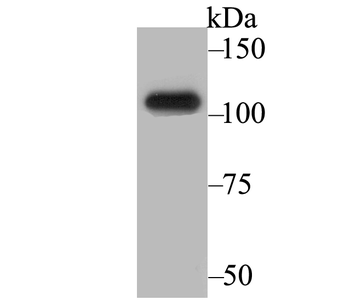
Western blot analysis of BRD2 on SiHa cell lysates. Proteins were transferred to a PVDF membrane and blocked with 5% BSA in PBS for 1 hour at room temperature. The primary antibody (orb1499352, 1/500) was used in 5% BSA at room temperature for 2 hours. Goat Anti-Rabbit IgG - HRP Secondary Antibody at 1:200000 dilution was used for 1 hour at room temperature. Predicted band size: 88 kDa, Observed band size: 105 kDa.
Similar Products
- Item 1 of 1
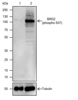
Phospho-BRD2 (Ser37) Recombinant Rabbit Monoclonal Antibody [orb1499303]
WB
Human
Rabbit
Recombinant
Unconjugated
25 μl, 100 μl, 50 μl 
Rabbit anti-BRD2 Recombinant Monoclonal Antibody [orb1519983]
FC, IP, WB
Human
Rabbit
Recombinant
Unconjugated
20 μlRabbit anti-BRD2 Recombinant Monoclonal Antibody [orb1519984]
FC, IP, WB
Human
Rabbit
Recombinant
Unconjugated
100 μl
Reviews
BRD2 Recombinant Rabbit Monoclonal Antibody (orb1499352)

- 0.0
Based on 0 reviews
Participating in our Biorbyt product reviews program enables you to support fellow scientists by sharing your firsthand experience with our products.
Login to Submit a Review