You have no items in your shopping cart.
Cart summary
Item 1 of 3
Item 1 of 3
BDNF Antibody: APC
Catalog Number: orb377476

| Catalog Number | orb377476 |
|---|---|
| Category | Antibodies |
| Description | Rabbit polyclonal antibody against BDNF conjugated to APC . |
| Species/Host | Rabbit |
| Clonality | Polyclonal |
| Tested applications | ICC, IF, IHC |
| Reactivity | Human, Mouse |
| Immunogen | Synthetic peptide from the N-terminal of human BDNF |
| Concentration | 1 mg/ml |
| Dilution range | WB (1:1000); ICC/IF (1:100) |
| Conjugation | APC |
| MW | 27.9kDa |
| Target | BDNF |
| Entrez | 627 |
| UniProt ID | P23560 |
| NCBI | NP_001137277.1 |
| Storage | Conjugated antibodies should be stored according to the product label |
| Buffer/Preservatives | 95.64mM Phosphate, 2.48mM MES and 2mM EDTA |
| Alternative names | Abrineurin antibody, ANON2 antibody, BDNF_Human an Read more... |
| Note | For research use only |
| Application notes | A 1:1000 dilution of SPC-703 was sufficient for detection of BDNF on 293T Rapamycin-treated lysates using Goat anti-rabbit IgG:HRP as the secondary antibody. |
| Expiration Date | 12 months from date of receipt. |

Immunocytochemistry/Immunofluorescence analysis using Rabbit Anti-BDNF Polyclonal Antibody. Tissue: Neuroblastoma cell line (SK-N-BE). Species: Human. Fixation: 4% Formaldehyde for 15 min at RT. Primary Antibody: Rabbit Anti-BDNF Polyclonal Antibody at 1:100 for 60 min at RT. Secondary Antibody: Goat Anti-Rabbit ATTO 488 at 1:100 for 60 min at RT. Counterstain: Phalloidin Texas Red F-Actin stain; DAPI (blue) nuclear stain at 1:1000, 1:5000 for 60min RT, 5min RT. Localization: Secreted, Cytoplasm, Membrane-bound vesicle. Magnification: 60X. (A) DAPI (blue) nuclear stain (B) Phalloidin Texas Red F-Actin stain (C) BDNF Antibody (D) Composite.
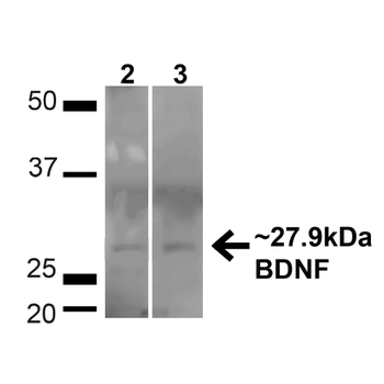
Western blot analysis of Human HeLa and HEK293T cell lysates showing detection of ~27.9 Kda BDNF protein using Rabbit Anti-BDNF Polyclonal Antibody. Lane 1: MW Ladder. Lane 2: Human HeLa (20 μg). Lane 3: Human 293T (20 μg). Load: 20 μg. Block: 5% milk + TBST for 1 hour at RT. Primary Antibody: Rabbit Anti-BDNF Polyclonal Antibody at 1:1000 for 1 hour at RT. Secondary Antibody: Goat Anti-Rabbit: HRP at 1:2000 for 1 hour at RT. Color Development: TMB solution for 12 min at RT. Predicted/Observed Size: ~27.9 Kda.
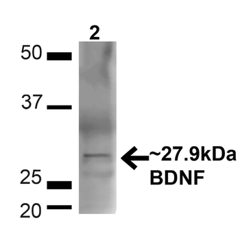
Western blot analysis of Mouse Brain showing detection of ~27.9 Kda BDNF protein using Rabbit Anti-BDNF Polyclonal Antibody. Lane 1: MW Ladder. Lane 2: Mouse Brain (20 μg). Load: 20 μg. Block: 5% milk + TBST for 1 hour at RT. Primary Antibody: Rabbit Anti-BDNF Polyclonal Antibody at 1:1000 for 1 hour at RT. Secondary Antibody: Goat Anti-Rabbit: HRP at 1:2000 for 1 hour at RT. Color Development: TMB solution for 12 min at RT. Predicted/Observed Size: ~27.9 Kda.
- Item 1 of 1

Phospho-TrkB (Tyr817) Rabbit Polyclonal Antibody (APC) [orb1006860]
IF
Human
Mouse, Rat
Rabbit
Polyclonal
APC
100 μlPhospho-TrkB (Tyr705) Rabbit Polyclonal Antibody (APC) [orb1007441]
ICC, IF
Bovine, Canine, Equine, Gallus, Porcine, Rabbit
Human, Mouse, Rat
Rabbit
Polyclonal
APC
100 μlPhospho-TrkB (Tyr515) Rabbit Polyclonal Antibody (APC) [orb1007442]
IF
Canine, Equine, Gallus, Porcine, Rabbit
Human, Mouse, Rat
Rabbit
Polyclonal
APC
100 μlBDNF Rabbit Polyclonal Antibody (APC) [orb1005283]
IF
Bovine, Canine, Equine, Guinea pig, Porcine, Rabbit
Human, Mouse, Rat
Rabbit
Polyclonal
APC
100 μl