You have no items in your shopping cart.
Cart summary
Item 1 of 5
Item 1 of 5
Basic Cytokeratin Antibody (HMW / Type II)
Catalog Number: orb248630

Product Properties
| Catalog Number | orb248630 |
|---|---|
| Category | Antibodies |
| Description | This antibody recognizes HMW Keratin 1, 5, 10 and 14. In normal epithelia, it stains stratified epithelia, myoepithelial cells and basal cells in the prostate gland and bronchi. The HMW Cytokeratin antibody shows no reactivity with hepatocytes, pancreatic acinar cells, proximal renal tubules, or endometrial glands; there is no reactivity with cells derived from simple epithelia. Mesenchymal tumors, lymphomas, melanomas, neural tumors, and neuroendocrine tumors are negative with this mAb. It stains myoepithelial cells and has been shown to be useful in distinguishing prostate adenocarcinoma from benign prostate. It has also been useful in separating benign from malignant intraductal breast proliferations. |
| Clonality | Monoclonal |
| Species/Host | Mouse |
| Isotype | Mouse IgG1, kappa |
| Conjugation | Unconjugated |
| Reactivity | Human, Mouse, Rat |
| Immunogen | Solubilized keratin extract from human stratum corneum was used as the immunogen for this HMW Cytokeratin antibody. |
| Tested applications | FACS, IF, IHC-P, WB |
| Dilution range | Flow cytometry: 1-2ug/million cells,Immunofluorescence: 1-2ug/ml,Western blot: 1-2ug/ml,Immunohistochemistry (FFPE): 0.25-0.5ug/ml for 30 min at RT |
| Application notes | The concentration stated for each application is a general starting point. Variations in protocols, secondaries and substrates may require the HMW Cytokeratin antibody to be titered up or down for optimal performance.1. Staining of formalin-fixed tissues requires boiling tissue sections in 10mM citrate buffer, pH 6.0, for 10-20 min followed by cooling at RT for 20 minutes.2. The prediluted format is supplied in a dropper bottle and is optimized for use in IHC. After epitope retrieval step (if required), drip mAb solution onto the tissue section and incubate at RT for 30 min. |
| Antibody Type | Primary Antibody |
| Clone Number | 34BE12 |
| Formula | 0.2 mg/ml in 1X PBS with 0.1 mg/ml BSA (US sourced) and 0.05% sodium azide |
| Storage | Maintain refrigerated at 2-8°C for up to 2 weeks. For long term storage store at -20°C in small aliquots to prevent freeze-thaw cycles. |
| Note | For research use only |
Images

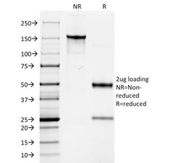
Western blot testing of human MCF7 cell lysate with HMW Cytokeratin antibody (clone 34BE12).
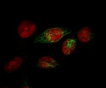
Immunofluorescent staining of MeOH-fixed human HeLa cells with HMW Cytokeratin antibody (clone 34BE12, green) and Reddot nuclear stain (red).
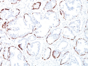
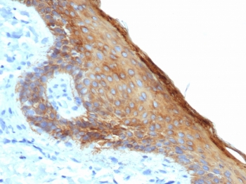
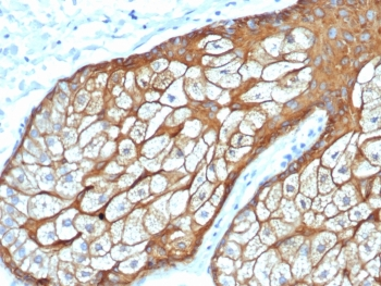
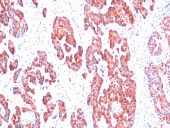
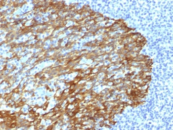
IHC staining of FFPE human prostate tissue with HMW Cytokeratin antibody (clone 34BE12). HIER: boil tissue sections in pH9 10mM Tris with 1mM EDTA for 20 min and allow to cool before testing.
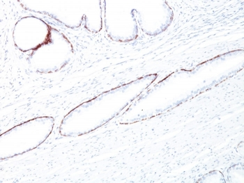
IHC staining of FFPE human prostate tissue with HMW Cytokeratin antibody (clone 34BE12). HIER: boil tissue sections in pH9 10mM Tris with 1mM EDTA for 20 min and allow to cool before testing.
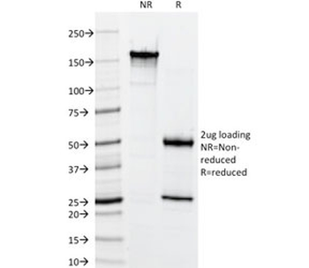
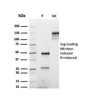
SDS-PAGE analysis of purified, BSA-free HMW Cytokeratin antibody (clone 34BE12) as confirmation of integrity and purity.
Similar Products
- Item 1 of 3
Basic Cytokeratin Antibody (HMW / Type II) [orb1151537]
IHC-P
Human
Mouse
Recombinant
Unconjugated
20 μg, 100 μg - Item 1 of 2
Basic Cytokeratin Antibody (HMW / Type II) [orb639458]
FACS, IF, IHC-P, WB
Human, Mouse, Rat
Mouse
Monoclonal
Unconjugated
20 μg, 100 μg - Item 1 of 1
Basic Cytokeratin Antibody (HMW / Type II) [orb606884]
IHC-P
Human
Mouse
Monoclonal
Unconjugated
20 μg, 100 μg - Item 1 of 1
Basic Cytokeratin Antibody (HMW / Type II) [orb640112]
IF, IHC-P
Human
Mouse
Monoclonal
Unconjugated
20 μg, 100 μg - Item 1 of 1
Basic Cytokeratin Antibody (HMW / Type II) [orb1825733]
IHC-P
Human
Mouse
Recombinant
Unconjugated
100 μg


Reviews
Basic Cytokeratin Antibody (HMW / Type II) (orb248630)

- 0.0
Based on 0 reviews
Participating in our Biorbyt product reviews program enables you to support fellow scientists by sharing your firsthand experience with our products.
Login to Submit a Review