You have no items in your shopping cart.
Cart summary
Item 1 of 4
Item 1 of 4
ATG9A Antibody (C-term)
Catalog Number: orb1933423

| Catalog Number | orb1933423 |
|---|---|
| Category | Antibodies |
| Description | Purified Rabbit Polyclonal Antibody (Pab) |
| Species/Host | Rabbit |
| Clonality | Polyclonal |
| Clone Number | RB7505, RB7506 |
| Tested applications | IF, IHC-P, WB |
| Reactivity | Human |
| Isotype | Rabbit IgG |
| Antibody Type | Primary Antibody |
| Dilution range | IF: 1:100, WB: 1:1000, WB: 1:1000, IHC-P: 1:50~100 |
| Form/Appearance | Purified polyclonal antibody supplied in PBS with 0.09% (W/V) sodium azide. This antibody is prepared by Saturated Ammonium Sulfate (SAS) precipitation followed by dialysis against PBS. |
| Conjugation | Unconjugated |
| MW | 94447 Da |
| Target | This ATG9A antibody is generated from rabbits immunized with a KLH conjugated synthetic peptide between 717-746 amino acids from the C-terminal region of human ATG9A. |
| UniProt ID | Q7Z3C6 |
| NCBI | NP_076990.4, NP_001070666.1 |
| Storage | Maintain refrigerated at 2-8°C for up to 2 weeks. For long term storage store at -20°C in small aliquots to prevent freeze-thaw cycles |
| Alternative names | Autophagy-related protein 9A, APG9-like 1, mATG9, Read more... |
| Note | For research use only |
| Expiration Date | 12 months from date of receipt. |
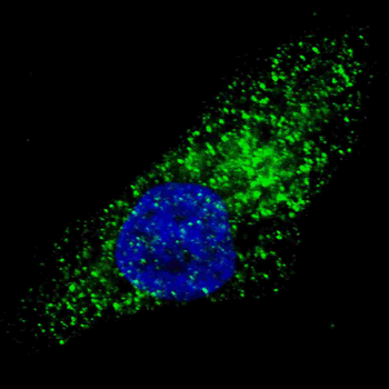
Fluorescent image of U251 cells stained with ATG9A (C-term) antibody. U251 cells were treated with Chloroquine (50 μM, 16 h), then fixed with 4% PFA (20 min), permeabilized with Triton X-100 (0.2%, 30 min). Cells were then incubated with ATG9A (C-term) primary antibody (1:100, 2 h at room temperature). For secondary antibody, Alexa Fluor 488 conjugated donkey anti-rabbit antibody (green) was used (1:1000, 1 h). Nuclei were counterstained with Hoechst 33342 (blue) (10 μg/ml, 5 min). ATG9A immunoreactivity is localized to autophagic vacuoles in the cytoplasm of U251 cells.
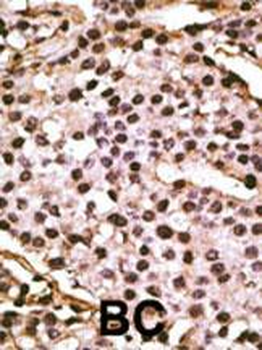
Formalin-fixed and paraffin-embedded human cancer tissue reacted with the primary antibody, which was peroxidase-conjugated to the secondary antibody, followed by DAB staining. This data demonstrates the use of this antibody for immunohistochemistry; clinical relevance has not been evaluated. BC = breast carcinoma; HC = hepatocarcinoma.

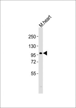
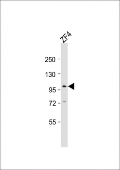
Western blot analysis of anti-Autophagy APG9L1 Antibody (C-term) in A2058 and A375 cell line lysates (35 ug/lane). APG9L1 (arrow) was detected using the purified Pab.
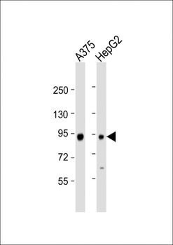
All lanes: Anti-APG9L1 Antibody (R732) at 1:1000 dilution. Lane 1: A375 whole cell lysate. Lane 2: HepG2 whole cell lysate. Lysates/proteins at 20 µg per lane. Secondary Goat Anti-Rabbit IgG, (H+L), Peroxidase conjugated at 1/10000 dilution. Predicted band size: 94 kDa. Blocking/Dilution buffer: 5% NFDM/TBST.
- Item 1 of 1
- Item 1 of 1