You have no items in your shopping cart.
Cart summary
Item 1 of 4
Item 1 of 4
CD5 Antibody
Catalog Number: orb154398

Product Properties
| Catalog Number | orb154398 |
|---|---|
| Category | Antibodies |
| Description | Mouse monoclonal antibody to CD5 which also designated Lyt-1 that has been identified as a transmembrane glycoprotein. It plays role as a receptor in regulating T-cell proliferation. CD5 interacts with CD72/LYB-2, as well as it is expressed at various developmental and activation stages on human B cells. |
| Target | CD5 |
| Clonality | Monoclonal |
| Isotype | Mouse IgG2a kappa |
| Conjugation | Unconjugated |
| Reactivity | Human |
| Concentration | 1 mg/ml |
| Buffer/Preservatives | Phosphate buffered saline (PBS), pH 7.4, 15 mM sodium azide |
| Purification | Purified by protein-A affinity chromatography. |
| Immunogen | Human acute lymphoblastic leukemia (ALL) T cells |
| UniProt ID | P06127 |
| Tested applications | FC, ICC, IHC, IP, WB |
| Application notes | Flow cytometry: Recommended dilution: 1-4 μg/ml.Western blotting: Laurylmaltoside lysing buffer; non-reducing conditions; recommended dilution: 1-2 μg/ml. |
| Antibody Type | Primary Antibody |
| Clone Number | L17F12 |
| Storage | Maintain refrigerated at 2-8°C for up to 2 weeks. For long term storage store at -20°C in small aliquots to prevent freeze-thaw cycles. |
| Alternative names | T1, LEU1 |
| Research Area | Epigenetics, Stem Cells |
| Note | For research use only |
| Entrez | 921 |
Images
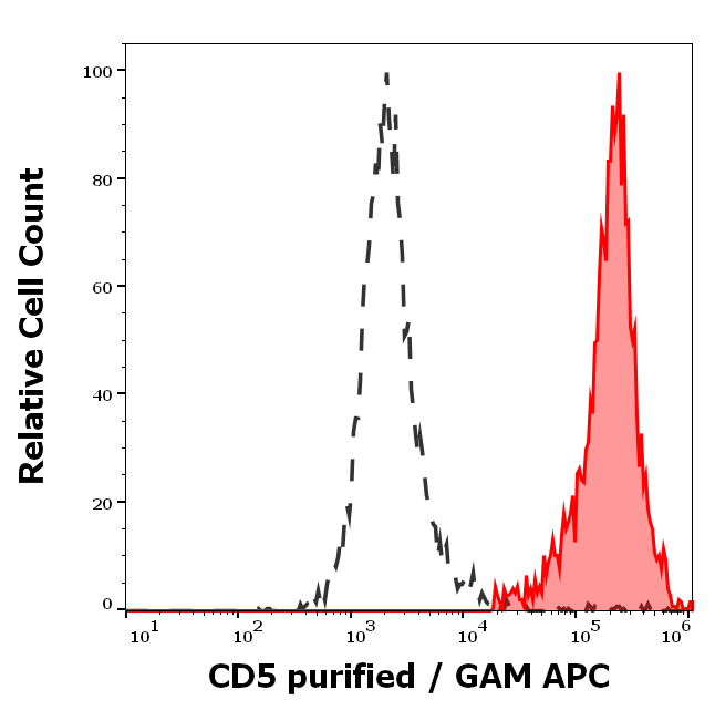
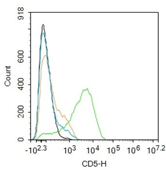
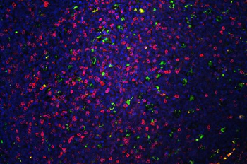
Separation of human CD5 positive lymphocytes (red-filled) from neutrophil granulocytes (black-dashed) in flow cytometry analysis (surface staining) of human peripheral whole blood stained using anti-human CD5 (L17F12) purified antibody (concentration in sample 2 μg/ml, GAM APC).
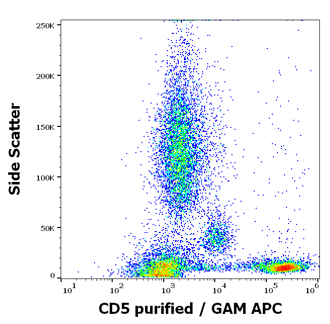
Flow cytometry surface staining pattern of human peripheral whole blood stained using anti-human CD5 (L17F12) purified antibody (concentration in sample 2 μg/ml, GAM APC).
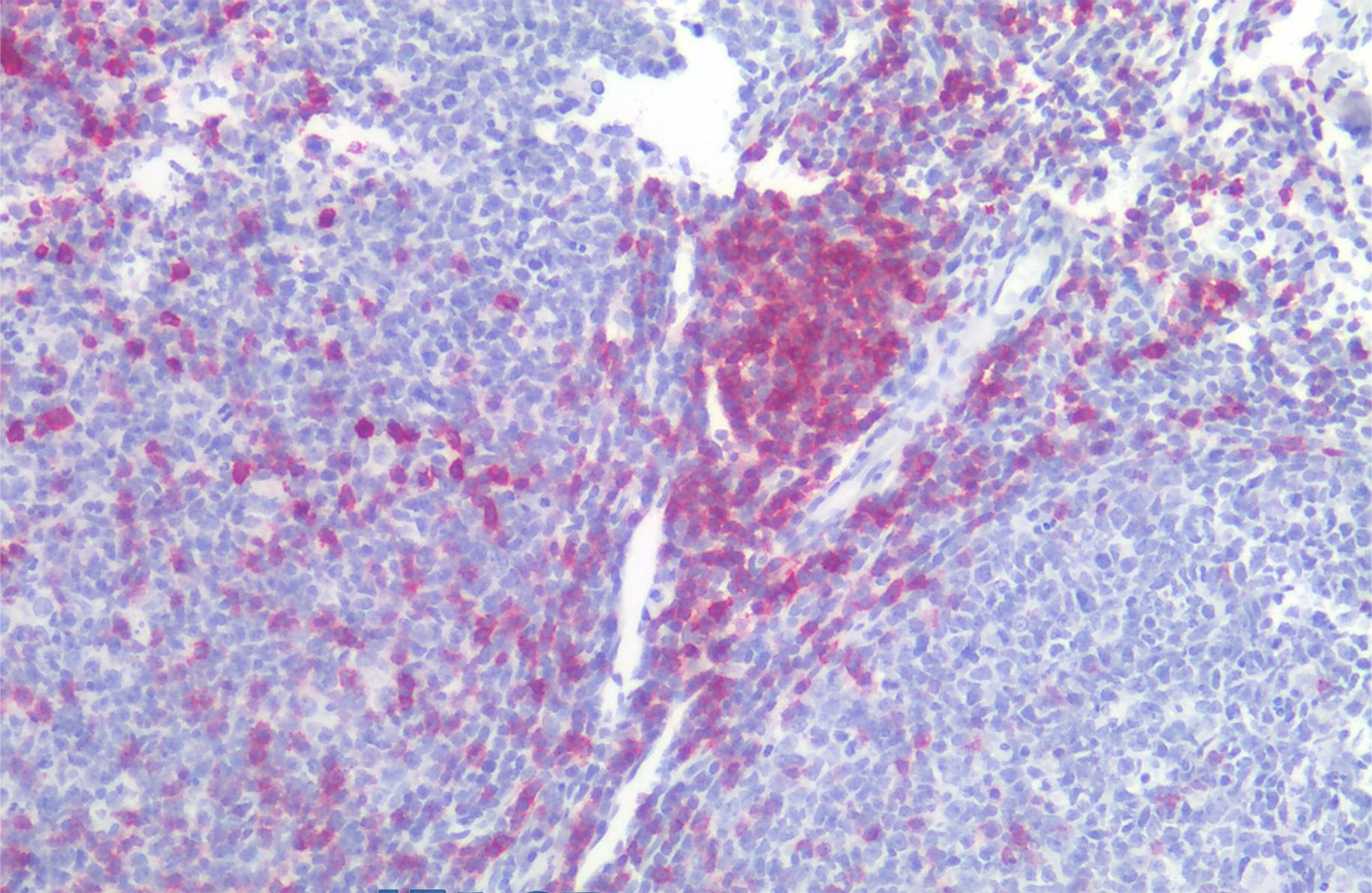
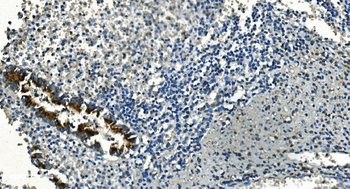
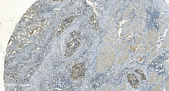
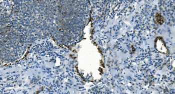
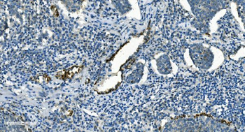
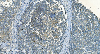
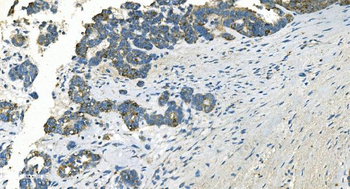
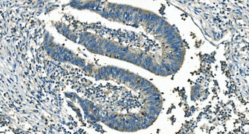
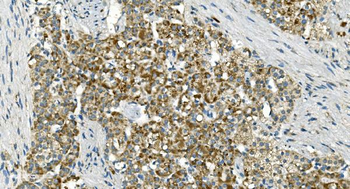
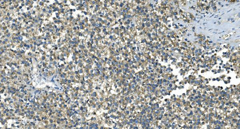
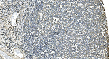
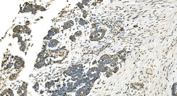
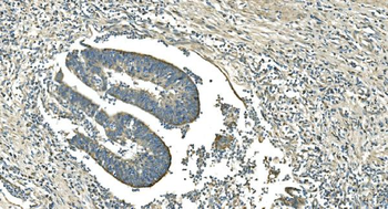
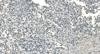
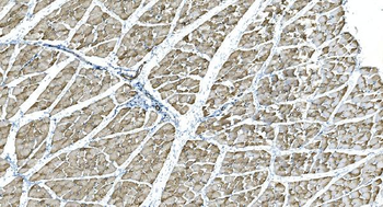
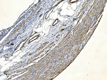
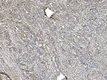
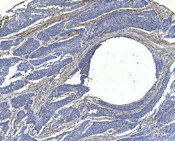
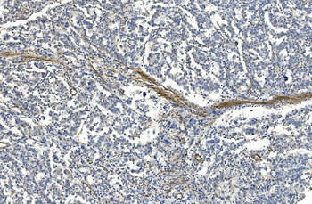
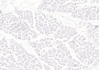
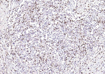
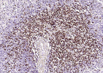
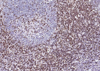
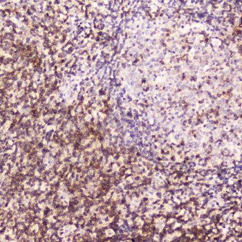
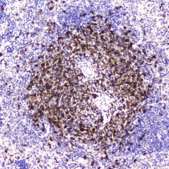
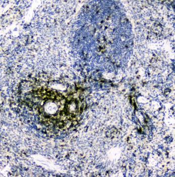
Immunohistochemistry staining of human tonsil (paraffin-embedded sections) with anti-CD5 (L17F12), 10 μg/ml.
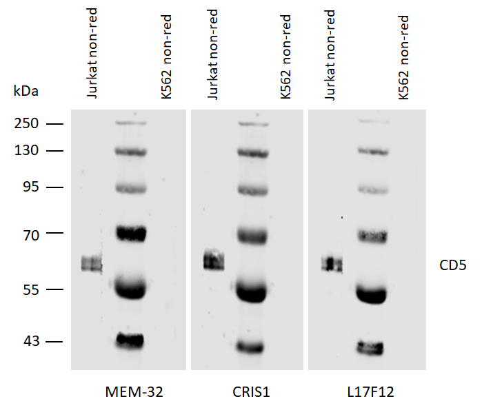
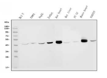
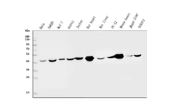
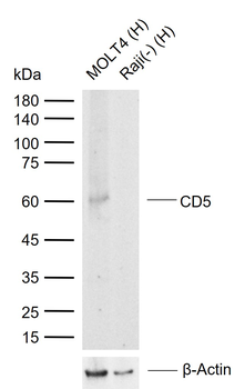
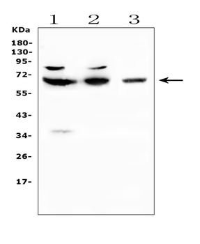
Western blotting analysis of human CD5 using mouse monoclonal antibodies MEM-32, CRIS1, and L17F12 on laurylmaltoside lysates of Jurkat cells and of K562 cells (negative control) under non-reducing conditions. Nitrocellulose membrane was probed with 2 µg/ml of mouse anti-CD5 monoclonal antibody followed by IRDye800-conjugated anti-mouse secondary antibody. CD5 was detected at approximately 62 kDa.
Similar Products
- Item 1 of 10
IDH2 Antibody (monoclonal, 2H4) [orb692220]
IHC, WB
Human, Mouse, Rat
Mouse
Monoclonal
Unconjugated
100 μg - Item 1 of 8
IDH2 Antibody (monoclonal, 6B13) [orb692221]
FC, ICC, IF, IHC, WB
Human, Mouse, Rat
Mouse
Monoclonal
Unconjugated
100 μg - Item 1 of 8
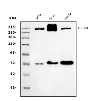
Filamin A/FLNA Antibody (monoclonal, 3F8) [orb692222]
IF, IHC, WB
Human
Mouse
Monoclonal
Unconjugated
100 μg - Item 1 of 6
CD5 Mouse Monoclonal Antibody [orb1595750]
FC, IF, IHC-Fr, IHC-P, WB
Mouse, Rat
Human
Mouse
Monoclonal
Unconjugated
50 μl, 100 μl, 200 μl - Item 1 of 6


Reviews
CD5 Antibody (orb154398)

- 0.0
Based on 0 reviews
Participating in our Biorbyt product reviews program enables you to support fellow scientists by sharing your firsthand experience with our products.
Login to Submit a Review