You have no items in your shopping cart.
Cart summary
Item 1 of 2
Item 1 of 2
Anti-Hu CD222 Biotin
Catalog Number: orb402816

| Catalog Number | orb402816 |
|---|---|
| Category | Antibodies |
| Description | Mouse monoclonal antibody against CD222 conjugated to biotin |
| Clonality | Monoclonal |
| Clone Number | MEM-238 |
| Tested applications | FC, IP, WB |
| Reactivity | Human, Primate |
| Isotype | Mouse IgG1 |
| Immunogen | Recombinant Vaccinia virus encoding CD222. |
| Antibody Type | Primary Antibody |
| Concentration | 1 mg/ml |
| Dilution range | Flow cytometry: Extracellular and intracellular staining; recommended dilution: 2-6 μg/ml. Western blotting: Recommended dilution: 1 µg/ml; non-reducing conditions. |
| Purity | Purified antibody is conjugated with biotin LC-NHS ester under optimum conditions and unconjugated antibody and free biotin are removed by size-exclusion chromatography. |
| Conjugation | Biotin |
| Target | CD222 |
| Entrez | 3482 |
| UniProt ID | P11717 |
| Storage | Store at 2-8°C. Do not freeze. |
| Buffer/Preservatives | Phosphate buffered saline (PBS), pH 7.4, 15 mM sodium azide |
| Alternative names | Anti-IGF2R antibody, anti-MPR1 antibody, anti-CIMP Read more... |
| Note | For research use only |
| Application notes | Flow cytometry: Extracellular and intracellular staining; recommended dilution: 2-6 μg/ml.Western blotting: Recommended dilution: 1 µg/ml; non-reducing conditions. |
| Expiration Date | 12 months from date of receipt. |
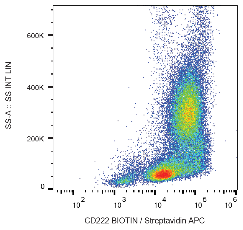
Flow cytometry analysis (surface staining) of human peripheral blood with anti-CD222 (MEM-238) biotin, streptavidin-APC.
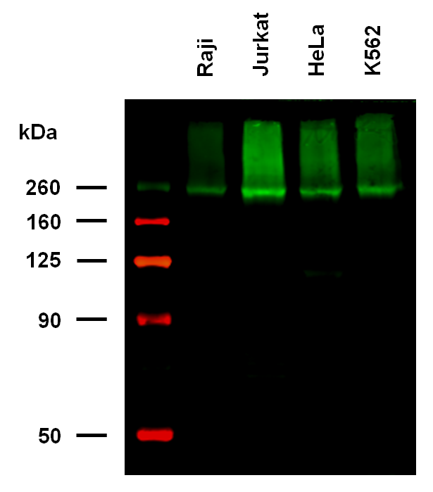
Anti-Hu CD222 Biotin (clone MEM-238) works in WB application under non-reducing conditions. Western blotting analysis was performed on whole cell extracts (RIPA lysis buffer) of Raji, Jurkat, HeLa, and K562 cell lines, mixed and heated (100°C, 5 min) with non-reducing SDS-loading buffer. Samples were resolved using 7% Tris-glycine SDS gel electrophoresis. Nitrocellulose membrane blot was probed with biotinylated mouse IgG1 monoclonal antibody MEM-238 (1 µg/ml), followed by IRDye 800CW Streptavidin (green). Multiplex fluorescent Western blot detection was performed. CD222 molecules were detected at ~250 kDa in all analysed cell lines.
