You have no items in your shopping cart.
Cart summary
Item 1 of 4
Item 1 of 4
Anti-Hu CD16 Purified
Catalog Number: orb44653

| Catalog Number | orb44653 |
|---|---|
| Category | Antibodies |
| Description | Mouse Monoclonal to CD16. |
| Target | CD16 |
| Clonality | Monoclonal |
| Isotype | Mouse IgG1 kappa |
| Conjugation | Unconjugated |
| Reactivity | Human, Primate |
| Concentration | 1 mg/ml |
| Buffer/Preservatives | Phosphate buffered saline (PBS), pH 7.4, 15 mM sodium azide |
| Purity | Purified by protein-A affinity chromatography. |
| Purification | Purified by protein-A affinity chromatography. |
| Immunogen | Human neutrophils |
| RRID | AB_10990854 |
| Tested applications | FC, IHC-Fr, IP |
| Application notes | Flow cytometry: Recommended dilution: 6 μg/ml. Immunohistochemistry (frozen sections): Acetone fixation. |
| Antibody Type | Primary Antibody |
| Clone Number | 3G8 |
| Storage | Maintain refrigerated at 2-8°C for up to 2 weeks. For long term storage store at -20°C in small aliquots to prevent freeze-thaw cycles. |
| Alternative names | Anti-CD16 antibody, anti-FACSgammaRIII antibody |
| Note | For research use only |
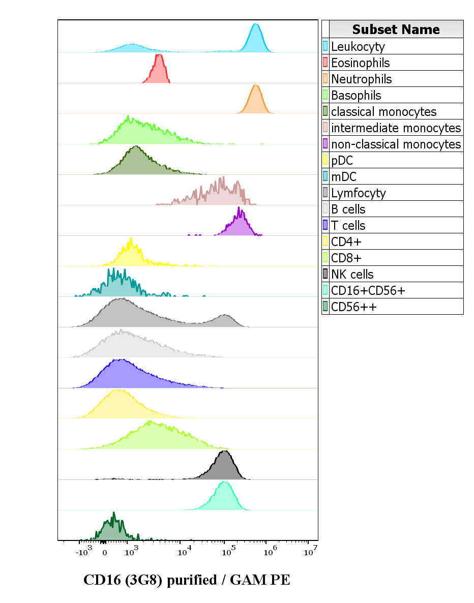
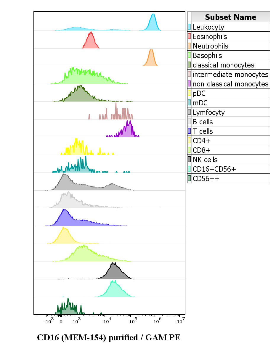
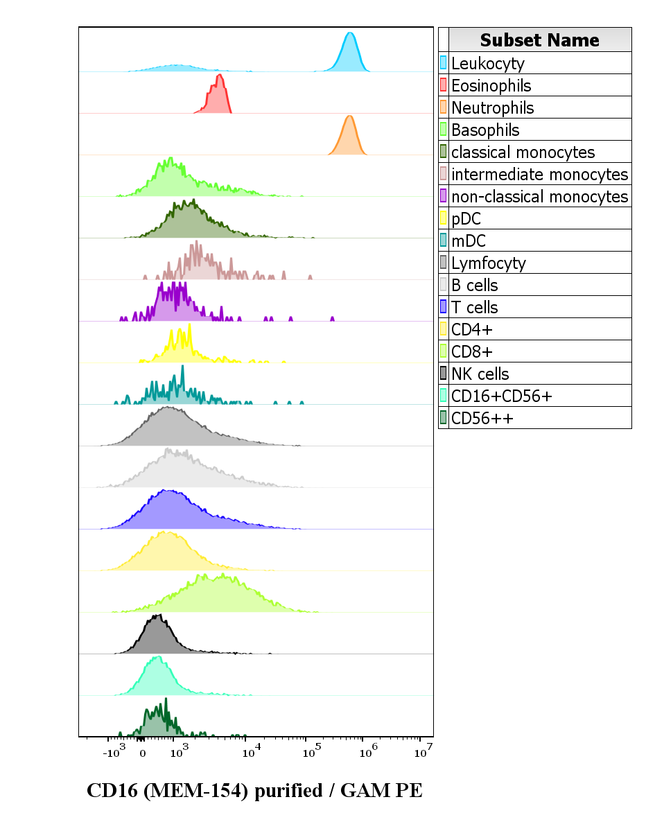
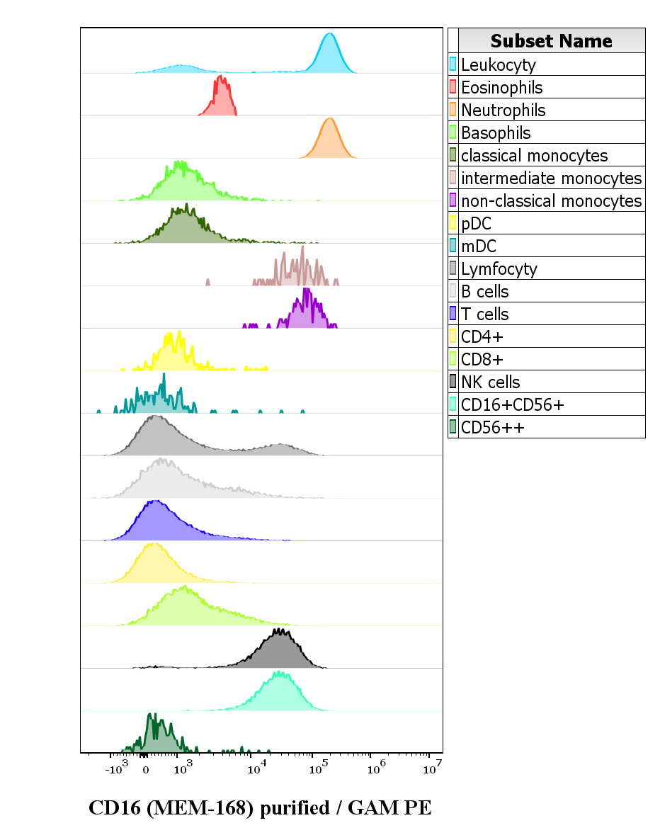
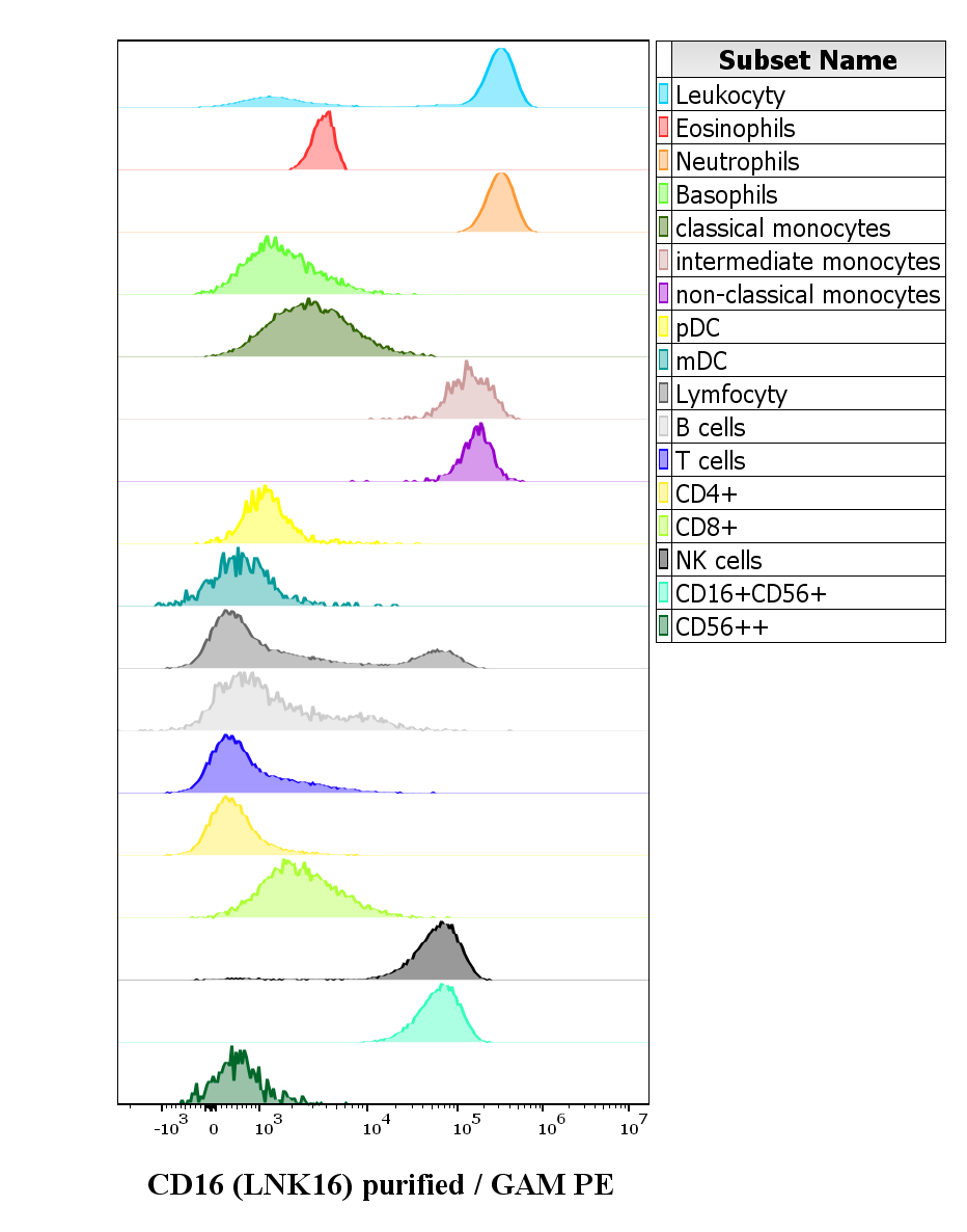
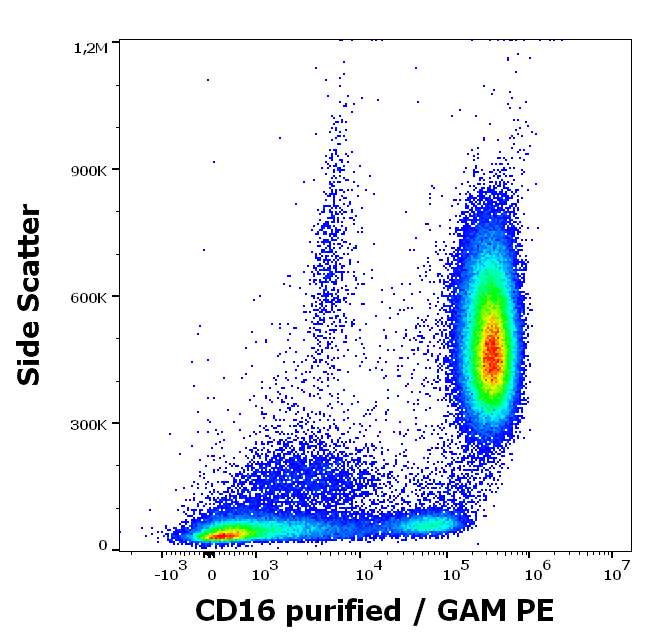
Expression profiling on peripheral blood subsets using anti-human CD16 purified antibody (clone 3G8). HCDM CDMaps standardized procedures were used for cell isolation and surface staining of blood leukocytes, with the modification of staining protocol using cytometry test tubes. Suspension of blood leukocytes isolated from buffy coats (2 x 10^6 cells) with residual erythrocytes lysed with 10× diluted EXCELLYSE Live solution was added to the mixture of anti-human CD16 purified antibody (clone 3G8, 0.5 µg/ml in stained blood sample) and Monocyte Blocking Buffer, vortexed and incubated for 20 min. Next, samples were centrifuged (670 g, 5 min.), supernatant removed and secondary antibody (GAM PE) was added to sample, vortexed and incubated for 20 min. Next, samples were washed twice (2 ml PBS, 670 g, 5 min.) and then optimized backbone antibody panels (HLDA Innate and HLDA Adaptive) were added to test tubes, vortexed and incubated for 20 min. Next, samples are fixed with 2 ml of 10× diluted EXCELLYSE Easy solution and incubated for 10 min. Finally, samples were centrifuged (670 g, 5 min.), supernatant removed and the cell pellet was resuspended in 200 µl of PBS for acquisition.
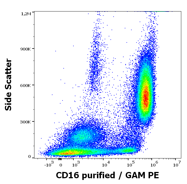
Anti-human CD16 purified antibody (clone 3G8) works in flow cytometry application. Analysis of the antibody staining profile was performed on blood leukocytes isolated from buffy coats. HCDM CDMaps standardized procedures were used for cell isolation and surface staining of blood leukocytes, with the modification of staining protocol using cytometry test tubes. Mouse monoclonal anti-human CD16 purified antibody (clone 3G8) was used in concentration 0.5 µg/ml in stained blood sample (2 x 10^6 cells).

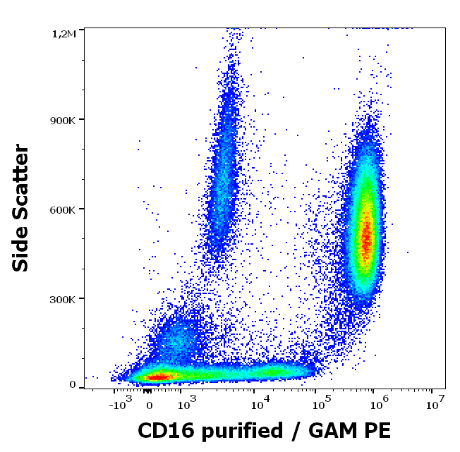
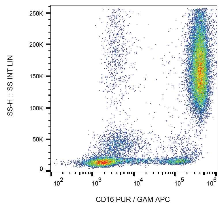
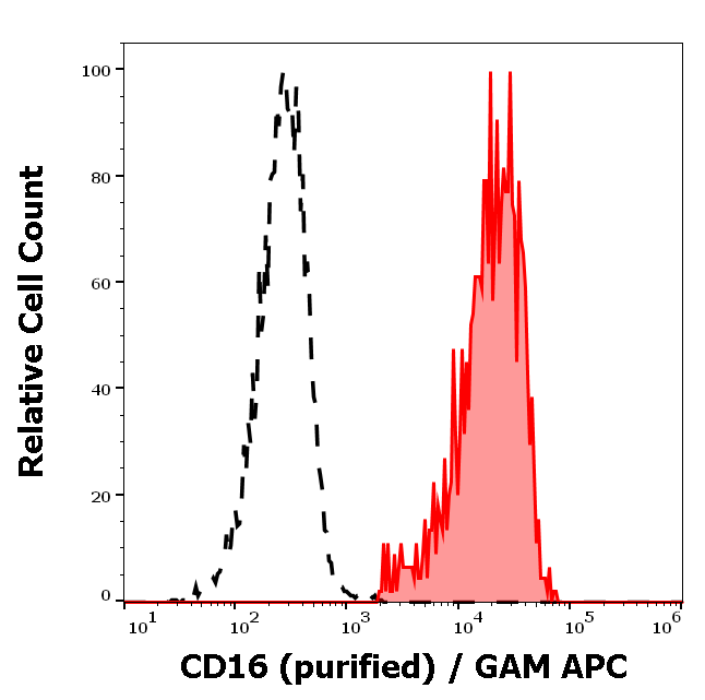
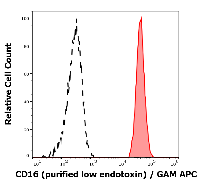
Separation of human CD16 positive lymphocytes (red-filled) from CD16 negative lymphocytes (black-dashed) in flow cytometry analysis (surface staining) of peripheral whole blood stained using anti-human CD16 (3G8) purified antibody (concentration in sample 2 µg/ml, GAM APC).

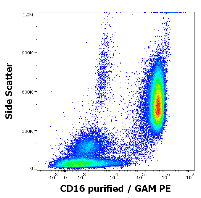
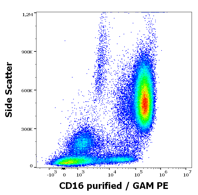
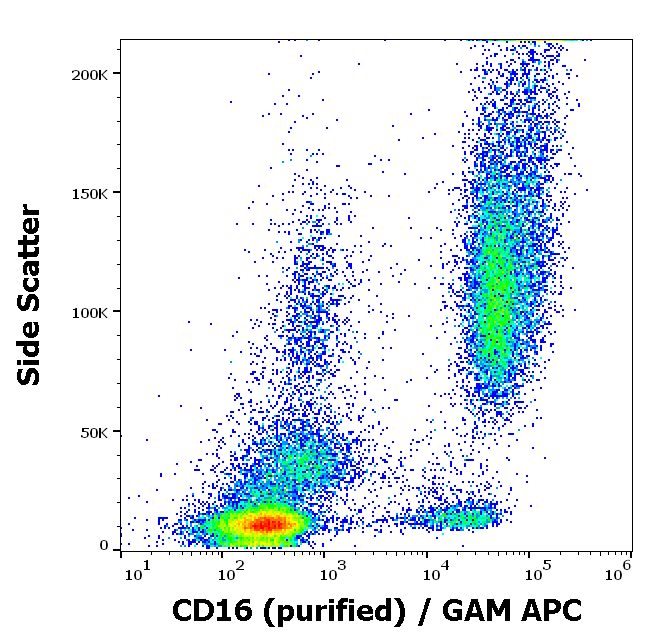
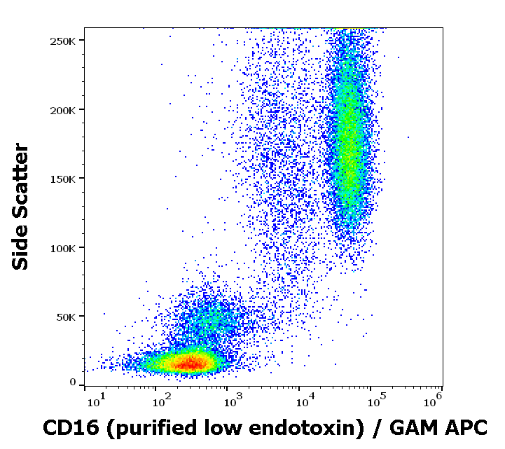
Flow cytometry surface staining pattern of human peripheral whole blood stained using anti-human CD16 (3G8) purified antibody (concentration in sample 2 µg/ml, GAM APC).
- Item 1 of 4
- Item 1 of 3
- Item 1 of 3
- Item 1 of 2
Anti-Hu CD16 Purified Low Endotoxin [orb154424]
FA, FC, IHC-Fr, IP
Human, Primate
Monoclonal
Unconjugated
0.1 mg - Item 1 of 2