You have no items in your shopping cart.
Cart summary
Item 1 of 3
Item 1 of 3

| Catalog Number | orb216142 |
|---|---|
| Category | Antibodies |
| Description | Rabbit polyclonal antibody to LGALS8 |
| Species/Host | Rabbit |
| Clonality | Polyclonal |
| Tested applications | IF, IH, WB |
| Reactivity | Human, Mouse |
| Immunogen | KLH-conjugated synthetic peptide encompassing a sequence within the center region of human Galectin 8. The exact sequence is proprietary. |
| Antibody Type | Primary Antibody |
| Dilution range | WB: 1:500-1:1000, IHC-P: 1:100-1-200, IF/ICC: 1:100-1:500 |
| Form/Appearance | Liquid in 0.42% Potassium phosphate, 0.87% Sodium chloride, pH 7.3, 30% glycerol, and 0.01% sodium azide. |
| Conjugation | Unconjugated |
| Target | LGALS8 |
| Entrez | 3964, 56048 |
| UniProt ID | O00214, Q9JL15 |
| Source | Rabbit |
| Storage | Maintain refrigerated at 2-8°C for up to 2 weeks. For long term storage store at -20°C in small aliquots to prevent freeze-thaw cycles. |
| Buffer/Preservatives | Liquid in 0.42% Potassium phosphate, 0.87% Sodium chloride, pH 7.3, 30% glycerol, and 0.01% sodium azide. |
| Alternative names | anti Galectin-8 antibody, anti Gal-8 antibody, ant Read more... |
| Note | For research use only |
| Expiration Date | 12 months from date of receipt. |
Filter by Applications
Filter by Reactivity
Yiting Wang 1, Yufan Sun 2, Shouyan Deng 1, Jiayang Liu 1, Jianghong Yu 1, Hao Chi 1, Xue Han 1, Yuan Zhang 1, Jiawei Shi 1, Yungang Wang 1, Yingfei Quan 2, Hai Li 3, Jie Xu 4 Discovery of galectin-8 as an LILRB4 ligand driving M-MDSCs defines a class of antibodies to fight solid tumors Cell Rep Med, 5, 101374
Applications
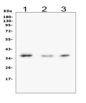
WB
Reactivity
Human
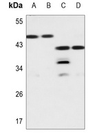
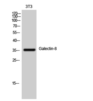
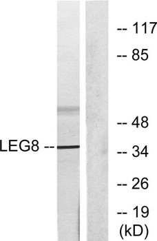
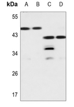
Western blot analysis of Galectin 8 expression in HEK293T (A), U2OS (B), mouse kidney (C), mouse testis (D) whole cell lysates. (Predicted band size: 35 kD; Observed band size: 38; 50 kD)
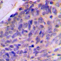
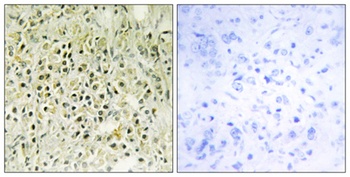
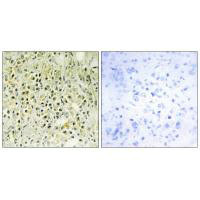
Immunohistochemical analysis of Galectin 8 staining in human breast cancer formalin fixed paraffin embedded tissue section. The section was pre-treated using heat mediated antigen retrieval with sodium citrate buffer (pH 6.0). The section was then incubated with the antibody at room temperature and detected using an HRP conjugated compact polymer system. DAB was used as the chromogen. The section was then counterstained with haematoxylin and mounted with DPX.
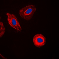
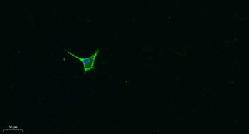
Immunofluorescent analysis of Galectin 8 staining in HepG2 cells. Formalin-fixed cells were permeabilized with 0.1% Triton X-100 in TBS for 5-10 minutes and blocked with 3% BSA-PBS for 30 minutes at room temperature. Cells were probed with the primary antibody in 3% BSA-PBS and incubated overnight at 4 °C in a humidified chamber. Cells were washed with PBST and incubated with a DyLight 594-conjugated secondary antibody (red) in PBS at room temperature in the dark. DAPI was used to stain the cell nuclei (blue).
- Item 1 of 4
Galectin-8 rabbit pAb [orb768984]
ELISA, IF, IHC-P, WB
Human, Mouse
Polyclonal
Unconjugated
50 μl, 100 μl - Item 1 of 3
Anti-Galectin 8 Antibody [orb1423069]
IF, IHC, WB
Human, Mouse
Rabbit
Polyclonal
Unconjugated
100 μl, 50 μl - Item 1 of 2
- Item 1 of 1
LGALS8 Antibody [orb672873]
ELISA, IF, IHC, WB
Human, Mouse
Rabbit
Polyclonal
Unconjugated
100 μg, 50 μg - Item 1 of 1
Anti-Galectin 8/LGALS8 Antibody [orb315167]
WB
Human, Mouse, Rat
Rabbit
Polyclonal
Unconjugated
10 μg, 100 μg