You have no items in your shopping cart.
Cart summary
Item 1 of 2
Item 1 of 2
Anti-Furin Antibody
Catalog Number: orb215936

| Catalog Number | orb215936 |
|---|---|
| Category | Antibodies |
| Description | Anti-Furin Antibody. Tested in WB applications. This antibody reacts with Human. |
| Species/Host | Rabbit |
| Clonality | Polyclonal |
| Tested applications | WB |
| Reactivity | Human |
| Isotype | Rabbit IgG |
| Immunogen | E.coli-derived human Furin recombinant protein (Position: T591-L794). Human Furin shares 88% amino acid (aa) sequence identity with both mouse and rat Furin. |
| Antibody Type | Primary Antibody |
| Concentration | Adding 0.2 ml of distilled water will yield a concentration of 500 μg/ml. |
| Form/Appearance | Lyophilized |
| Conjugation | Unconjugated |
| MW | 87 kDa |
| UniProt ID | P09958 |
| Storage | Maintain refrigerated at 2-8°C for up to 2 weeks. For long term storage store at -20°C in small aliquots to prevent freeze-thaw cycles. |
| Alternative names | Furin; 3.4.21.75; Dibasic-processing enzyme; Paire Read more... |
| Note | For research use only |
| Application notes | Western blot, 0.1-0.5μg/ml, Human. Add 0.2ml of distilled water will yield a concentration of 500ug/ml |
| Expiration Date | 12 months from date of receipt. |
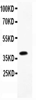
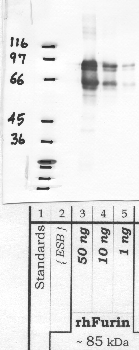
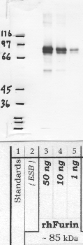
Western blot analysis of Furin using anti-Furin antibody. Electrophoresis was performed on a 5-20% SDS-PAGE gel at 70V (Stacking gel) / 90V (Resolving gel) for 2-3 hours. Lane 1: recombinant human Furin protein 0.5 ng. After electrophoresis, proteins were transferred to a nitrocellulose membrane at 150 mA for 50-90 minutes. Blocked the membrane with 5% non-fat milk/TBS for 1.5 hour at RT. The membrane was incubated with rabbit anti-Furin antigen affinity purified polyclonal antibody at 0.5 µg/mL overnight at 4°C, then washed with TBS-0.1% Tween 3 times with 5 minutes each and probed with a goat anti-rabbit IgG-HRP secondary antibody at a dilution of 1:5000 for 1.5 hour at RT. The signal is developed using an Enhanced Chemiluminescent detection (ECL) kit with Tanon 5200 system. A specific band was detected for Furin at approximately 40 kDa. The expected band size for Furin is at 40 kDa.
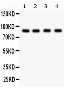
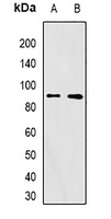
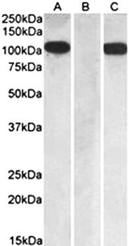
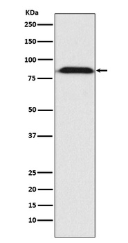
Western blot analysis of Furin using anti-Furin antibody. Electrophoresis was performed on a 5-20% SDS-PAGE gel at 70V (Stacking gel) / 90V (Resolving gel) for 2-3 hours. The sample well of each lane was loaded with 30 ug of sample under reducing conditions. Lane 1: human Hela whole cell lysates, Lane 2: human MCF-7 whole cell lysates, Lane 3: human Colo320 whole cell lysates, Lane 4: human SW620 whole cell lysates. After electrophoresis, proteins were transferred to a nitrocellulose membrane at 150 mA for 50-90 minutes. Blocked the membrane with 5% non-fat milk/TBS for 1.5 hour at RT. The membrane was incubated with rabbit anti-Furin antigen affinity purified polyclonal antibody at 0.5 µg/mL overnight at 4°C, then washed with TBS-0.1% Tween 3 times with 5 minutes each and probed with a goat anti-rabbit IgG-HRP secondary antibody at a dilution of 1:5000 for 1.5 hour at RT. The signal is developed using an Enhanced Chemiluminescent detection (ECL) kit with Tanon 5200 system. A specific band was detected for Furin at approximately 87 kDa. The expected band size for Furin is at 87 kDa.
- Item 1 of 1
- Item 1 of 1
- Item 1 of 3
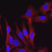
Anti-FURIN Antibody [orb378067]
IF, IH, WB
Human, Mouse, Rat
Rabbit
Polyclonal
Unconjugated
200 μl, 100 μl, 50 μl - Item 1 of 1
Goat anti-FURIN / PCSK3 Antibody [orb131685]
ELISA, IF
Human, Mouse, Rat
Goat
Polyclonal
Unconjugated
100 μg - Item 1 of 1
Anti-Furin Monoclonal Antibody [orb547715]
ICC, IF, IHC, WB
Human, Mouse, Rat
Rabbit
Monoclonal
Unconjugated