You have no items in your shopping cart.
Cart summary
Item 1 of 3
Item 1 of 3
EPHA1 Antibody
Catalog Number: orb318931

Product Properties
| Catalog Number | orb318931 |
|---|---|
| Category | Antibodies |
| Description | The EPHA1 Antibody is suitable for IF, IHC, WB. It is a Polyclonal, Unconjugated antibody which raised against KLH-conjugated synthetic peptide encompassing a sequence within the center region of human EPHA1. The exact sequence is proprietary. Purification: The antibody was purified by immunogen affinity chromatography. |
| Clonality | Polyclonal |
| Species/Host | Rabbit |
| Conjugation | Unconjugated |
| Reactivity | Human |
| Form/Appearance | Liquid in 0.42% Potassium phosphate, 0.87% Sodium chloride, pH 7.3, 30% glycerol, and 0.01% sodium azide. |
| Purification | The antibody was purified by immunogen affinity chromatography. |
| Immunogen | KLH-conjugated synthetic peptide encompassing a sequence within the center region of human EPHA1. The exact sequence is proprietary. |
| UniProt ID | P21709 |
| Tested applications | IF, IHC, WB |
| Dilution range | WB: 1:500-1:1000, IHC-P: 1:100-1:200, IF/ICC: 1:100-1:500 |
| Antibody Type | Primary Antibody |
| Storage | Maintain refrigerated at 2-8°C for up to 2 weeks. For long term storage store at -20°C in small aliquots to prevent freeze-thaw cycles. |
| Alternative names | EPH; EPHT; EPHT1; Ephrin type-A receptor 1; hEpha1 Read more... |
| Research Area | Cancer, Epigenetics |
| Note | For research use only |
| Entrez | 2041 |
| Expiration Date | 12 months from date of receipt. |
Images
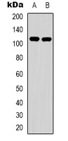
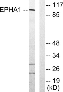
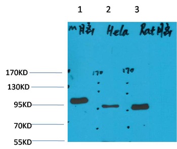
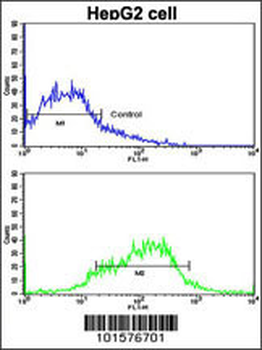
Western blot analysis of EPHA1 expression in Hela (A), HepG2 (B) whole cell lysates. (Predicted band size: 108 kD; Observed band size: 108 kD)
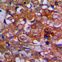
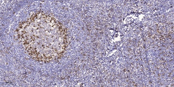
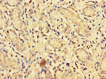
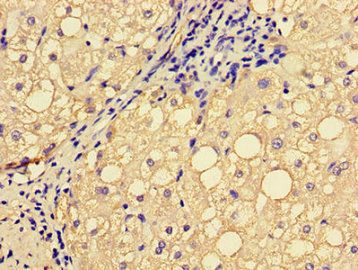
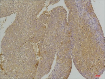
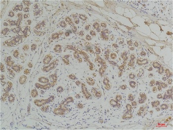
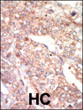
Immunohistochemical analysis of EPHA1 staining in human prostate cancer formalin fixed paraffin embedded tissue section. The section was pre-treated using heat mediated antigen retrieval with sodium citrate buffer (pH 6.0). The section was then incubated with the antibody at room temperature and detected using an HRP conjugated compact polymer system. DAB was used as the chromogen. The section was then counterstained with haematoxylin and mounted with DPX.
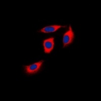
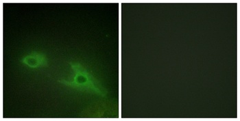
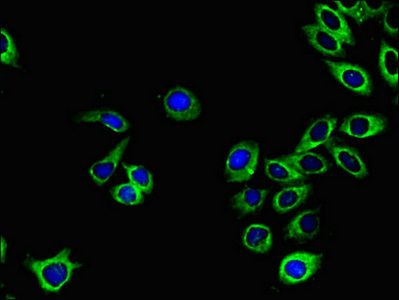
Immunofluorescent analysis of EPHA1 staining in Hela cells. Formalin-fixed cells were permeabilized with 0.1% Triton X-100 in TBS for 5-10 minutes and blocked with 3% BSA-PBS for 30 minutes at room temperature. Cells were probed with the primary antibody in 3% BSA-PBS and incubated overnight at 4 °C in a hidified chamber. Cells were washed with PBST and incubated with a DyLight 594-conjugated secondary antibody (red) in PBS at room temperature in the dark. DAPI was used to stain the cell nuclei (blue).
Similar Products
- Item 1 of 1
Human Ephrin Type A Receptor 1 (EPHA1) ELISA Kit [orb775763]
Human
0.32-20 ng/mL
0.121 ng/mL
48 T, 96 T - Item 1 of 4
EphA1 rabbit pAb [orb765145]
ELISA, IF, IHC-P, WB
Human, Mouse, Rat
Polyclonal
Unconjugated
100 μl, 50 μl - Item 1 of 4
- Item 1 of 3
- Item 1 of 3


Reviews
EPHA1 Antibody (orb318931)

- 0.0
Based on 0 reviews
Participating in our Biorbyt product reviews program enables you to support fellow scientists by sharing your firsthand experience with our products.
Login to Submit a Review