You have no items in your shopping cart.
Cart summary
Item 1 of 3
Item 1 of 3
Ankyrin G Antibody: FITC
Catalog Number: orb149896

| Catalog Number | orb149896 |
|---|---|
| Category | Antibodies |
| Description | Mouse monoclonal to Ankyrin G (FITC). Ankyrins are a family of adaptor proteins that mediate the attachment of integral membrane proteins to the spectrin-actin based membrane skeleton. Ankyrins have binding sites for the beta subunit of spectrin and at least 12 families of integral membrane proteins. This linkage is required to maintain the integrity of the plasma membranes and to anchor specific ion channels, ion exchangers and ion transporters in the plasma membrane.. |
| Species/Host | Mouse |
| Clonality | Monoclonal |
| Clone Number | N106/20 (Formerly sold as S106-20) |
| Tested applications | ICC, IF, IHC |
| Reactivity | Human, Mouse, Rat |
| Isotype | IgG1 |
| Immunogen | Fusion protein 1000 C-terminal amino acids of human Ankyrin G (also known as ANK-3 or ankyrin-3) encompassing all of Ankyrin G with the exception of Ankyrin repeats |
| Concentration | 1 mg/ml |
| Dilution range | WB (1:1000), ICC/IF (1:100) |
| Conjugation | FITC |
| MW | 200kDa |
| Target | Ankyrin G |
| Entrez | 288 |
| UniProt ID | Q12955 |
| NCBI | NP_066267.2 |
| Storage | Conjugated antibodies should be stored according to the product label |
| Buffer/Preservatives | 640.91mM DMSO, 136.36mM Ethanolamine, 9.09mM Sodium Bicarbonate in 90.9% PBS |
| Alternative names | ANK-3 antibody, ANK3 antibody, ankyrin 3 antibody, Read more... |
| Note | For research use only |
| Application notes | 1 µg/ml of SMC-404 was sufficient for detection of Ankyrin-G in 20 µg of rat brain membrane lysate and assayed by colorimetric immunoblot analysis using goat anti-mouse IgG:HRP as the secondary antibody. |
| Expiration Date | 12 months from date of receipt. |
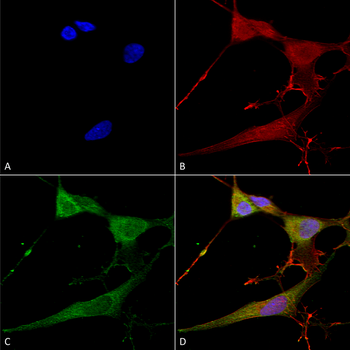
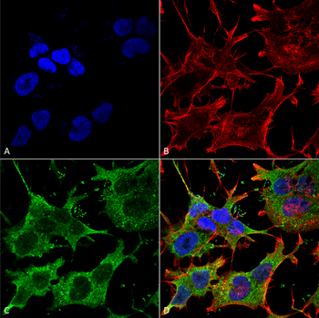
Immunocytochemistry/Immunofluorescence analysis using Mouse Anti-Ankyrin G Monoclonal Antibody, Clone N106/20. Tissue: Neuroblastoma cells (SH-SY5Y). Species: Human. Fixation: 4% PFA for 15 min. Primary Antibody: Mouse Anti-Ankyrin G Monoclonal Antibody at 1:100 for overnight at 4°C with slow rocking. Secondary Antibody: AlexaFluor 488 at 1:1000 for 1 hour at RT. Counterstain: Phalloidin-iFluor 647 (red) F-Actin stain; Hoechst (blue) nuclear stain at 1:800, 1.6mM for 20 min at RT. (A) Hoechst (blue) nuclear stain. (B) Phalloidin-iFluor 647 (red) F-Actin stain. (C) Ankyrin G Antibody (D) Composite.
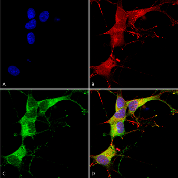
Immunocytochemistry/Immunofluorescence analysis using Mouse Anti-Ankyrin G Monoclonal Antibody, Clone N106/20. Tissue: Neuroblastoma cell line (SK-N-BE). Species: Human. Fixation: 4% Formaldehyde for 15 min at RT. Primary Antibody: Mouse Anti-Ankyrin G Monoclonal Antibody at 1:100 for 60 min at RT. Secondary Antibody: Goat Anti-Mouse ATTO 488 at 1:200 for 60 min at RT. Counterstain: Phalloidin Texas Red F-Actin stain; DAPI (blue) nuclear stain at 1:1000, 1:5000 for 60 min at RT, 5 min at RT. Localization: Cytoplasm. Magnification: 60X. (A) DAPI (blue) nuclear stain. (B) Phalloidin Texas Red F-Actin stain. (C) Ankyrin G Antibody. (D) Composite.
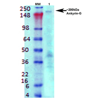
Western Blot analysis of Rat brain membrane lysate showing detection of Ankyrin G protein using Mouse Anti-Ankyrin G Monoclonal Antibody, Clone N106/20. Primary Antibody: Mouse Anti-Ankyrin G Monoclonal Antibody at 1:1000.
- Item 1 of 1

Ankyrin G Rabbit Polyclonal Antibody (FITC) [orb559573]
FC
Bovine, Canine, Equine, Mouse, Porcine, Rabbit, Rat
Human
Rabbit
Polyclonal
FITC
100 μl