You have no items in your shopping cart.
Cart summary
Item 1 of 5
Item 1 of 5
PIM2 antibody
Catalog Number: orb131688
| Catalog Number | orb131688 |
|---|---|
| Category | Antibodies |
| Description | Goat polyclonal to PIM2 |
| Species/Host | Goat |
| Clonality | Polyclonal |
| Tested applications | ELISA, IHC, WB |
| Reactivity | Human |
| Dilution range | ELISA: 1:16000, WB: 0.5-1 μg/ml, IHC-P: 5 μg/ml |
| Conjugation | Unconjugated |
| MW | 34.2 |
| Target | PIM2 |
| Entrez | 11040 |
| Protein Sequence | QTPAEDVPLNPSK |
| Storage | Aliquot and store at -20°C. Minimize freezing and thawing. |
| Buffer/Preservatives | Supplied at 0.5 mg/ml in Tris saline, 0.02% sodium azide, pH 7.3 with 0.5% bovine serum albumin. Aliquot and store at -20°C. Minimize freezing and thawing. |
| Alternative names | PIM2; pim-2 oncogene; pim-2h; proto-oncogene Pim-2 Read more... |
| Note | For research use only |
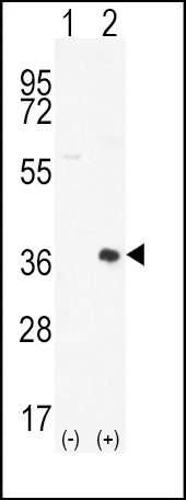
| Application notes | In transfected HEK293 transiently expressing Human PIM2 (myc and DYKDDDDK tagged), a band of approx. 38kDa is observed. No bands are observed in mock-transfected HEK293 and the same band is observed using anti-myc antibody. Recommended concentration, 0.5-1 μg/ml.WB: In transfected HEK293 transiently expressing Human PIM2 (myc and DYKDDDDK tagged), a band of approx. 38kDa is observed. No bands are observed in mock-transfected HEK293 and the same band is observed using anti-myc antibody. Recommended concentration, 0.5-1 μg/ml. |
| Expiration Date | 12 months from date of receipt. |
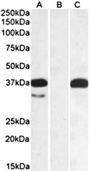
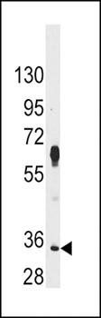
Western blot analysis of HEK293 lysate (10ug protein in RIPA buffer) overexpressing Human using PIM2 antibody
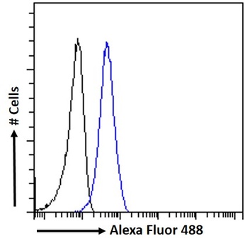
orb131688 Flow cytometric analysis of paraformaldehyde fixed HeLa cells (blue line), permeabilized with 0.5% Triton. Primary incubation 1hr (10ug/ml) followed by Alexa Fluor 488 secondary antibody (1ug/ml). IgG control: Unimmunized goat IgG (black line) followed by Alexa Fluor 488 secondary antibody.
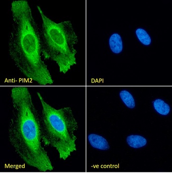
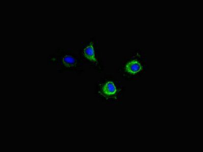
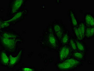
orb131688 Immunofluorescence analysis of paraformaldehyde fixed HeLa cells, permeabilized with 0.15% Triton. Primary incubation 1hr (10ug/ml) followed by Alexa Fluor 488 secondary antibody (2ug/ml), showing cytoplasmic and membrane staining. The nuclear stain is DAPI (blue). Negative control: Unimmunized goat IgG (10ug/ml) followed by Alexa Fluor 488 secondary antibody (2ug/ml).
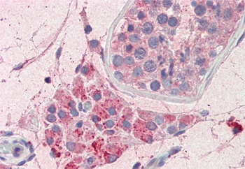
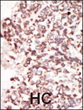
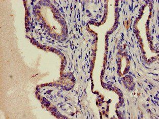
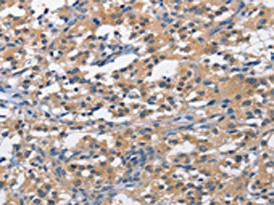
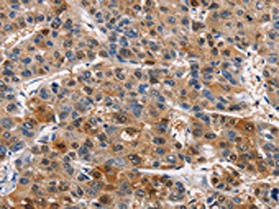
orb131688 (5µg/ml) staining of paraffin embedded Human Testis. Steamed antigen retrieval with citrate buffer pH 6, AP-staining.
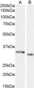
orb131688 (1µg/ml) staining of HepG2 (A) and K562 (B) cell lysate (35µg protein in RIPA buffer). Detected by chemiluminescence.
- Item 1 of 5
- Item 1 of 3
- Item 1 of 4
- Item 1 of 2
- Item 1 of 2


Submit a review
Filter by Rating

- 5 stars

- 4 stars
- 3 stars
- 2 stars
- 1 stars