
You have no items in your shopping cart.
Technical Guide & Reagents
Immunohistochemistry vs Immunocytochemistry
Understand the key differences between IHC and ICC, explore their roles in cancer diagnostics, and discover highly validated primary antibodies, secondary antibodies, and essential workflow reagents.
1. Technique Overview
Immunohistochemistry (IHC) and immunocytochemistry (ICC) are fundamental laboratory techniques used to identify particular protein antigens through the highly specific binding of antibodies.
Within these processes, a primary antibody attaches to the desired antigen. A secondary antibody, attached either to a fluorophore or to an enzyme (like HRP) whose catalytic process results in a colorimetric/fluorescent signal, then binds to the primary antibody. This allows researchers to precisely localize and identify target proteins within the sample.
2. Key Differences & Applications
While these methods have the same underlying immunological principles and overall process, there are a few critical differences regarding sample preparation, biological context, and downstream applications.
Immunohistochemistry (IHC)
- Sample Type: Utilizes whole tissue samples that are either snap-frozen or formalin-fixed and paraffin-embedded (FFPE).
- Biological Context: Because IHC utilizes intact tissue samples, researchers can study how cells interact within their native microenvironment and extracellular matrix.
- Applications: Widely used in clinical pathology, disease diagnosis, and cancer staging through the examination of tissue morphology and spatial protein expression.
Immunocytochemistry (ICC)
- Sample Type: Utilizes cultured cells, cell lines, or cell suspensions isolated from their extracellular matrix.
- Biological Context: Because ICC lacks complex tissue architecture, efforts are focused purely on isolated cell behavior and sub-cellular structures.
- Applications: Ideal for high-resolution imaging of cellular processes, precise intracellular protein localization (e.g., nuclear vs. cytoplasmic), and evaluating drug responses in vitro.
Cancer Research Impact: Both IHC and ICC have critical applications in oncology. IHC is essential for tumor diagnosis and staging by visualizing tumor markers across complex tissue biopsies. ICC is highly valuable for evaluating how isolated cancer cell lines respond to novel drug treatments, inducing apoptosis, or altering signaling pathways.
3. Primary Cancer Biomarkers for IHC & ICC
Biorbyt offers a wide array of highly specific primary antibodies optimized for IHC on tissue biopsies and ICC on cultured cancer cells. Below are some of the most critical diagnostic and structural markers.
| Biorbyt SKU | Target / Description | Clinical / Research Relevance | Example |
|---|---|---|---|
| orb1294317 | Anti-HER2 / ERBB2 AntibodyRecombinant mAb | Common biomarker for breast cancer and gastric cancer. | .png) |
| orb1274540 | Anti-EGFR AntibodyKO Validated mAb | Common biomarker for non-small cell lung cancer and glioblastoma. | 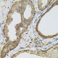 |
| orb69143 | Anti-BRAF AntibodyPrimary Antibody | Crucial mutation biomarker for melanoma and colorectal cancer. | 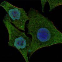 |
| orb53139 | Anti-KRAS AntibodyPrimary Antibody | Common biomarker for pancreatic and colorectal cancers. | 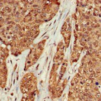 |
| orb556705 | Anti-PSA (PSAT1) AntibodyPrimary Antibody | Standard diagnostic and monitoring biomarker for prostate cancer. |  |
| orb247042 | Anti-Ki-67 AntibodyPrimary Antibody | Cellular marker strictly associated with active cell proliferation (tumor grading). | |
| orb11101 | Anti-E-Cadherin AntibodyPolyclonal Antibody | Epithelial marker; loss of E-Cadherin indicates metastasis and EMT. | |
| orb707372 | Anti-Caspase-3 AntibodyPrimary Antibody | Key executioner of apoptosis; evaluates drug response in cancer cells. |
4. Essential Workflow Reagents
Successful IHC and ICC staining requires more than just primary antibodies. Proper fixation, permeabilization, antigen retrieval, and signal detection are critical. Biorbyt provides the foundational buffers and substrates needed to perfect your workflow.
DAPI Nuclear Counterstain
Essential blue fluorescent stain that binds strongly to A-T rich regions in DNA. Perfect for defining nuclear boundaries in ICC/IHC.
View Products →Antigen Retrieval Buffers
Citrate and EDTA buffers designed to break protein cross-links formed during formalin fixation, unmasking epitopes for IHC.
View Buffers →DAB Substrate Kits
3,3'-Diaminobenzidine (DAB) produces a robust, insoluble brown precipitate when reacted with HRP, ideal for brightfield IHC.
View Kits →Normal Goat Serum
High-quality blocking buffer to prevent non-specific binding of secondary antibodies, ensuring a clean, low-background signal.
View Blocking Serums →Triton X-100
A non-ionic surfactant used for cellular permeabilization in ICC, allowing large antibodies to access intracellular targets.
View Permeabilizers →Mounting Media
Anti-fade fluoroshield mounting solutions to preserve tissue samples and protect fluorescent ICC signals from photobleaching.
View Mounting Media →5. Secondary Antibodies & Conjugates
Maximize your signal-to-noise ratio with our highly cross-adsorbed secondary antibodies. Available with enzymatic reporters (HRP, AP) for brightfield IHC or diverse fluorophores (FITC, Alexa Fluor equivalents) for multiplexed fluorescent ICC.
| Biorbyt SKU | Product Name | Conjugate Type | Target Species |
|---|---|---|---|
| orb3167248 | HRP Chicken Anti-Rabbit IgGSecondary Antibody | Enzymatic (HRP) | Rabbit IgG |
| orb3167245 | FITC Chicken Anti-Mouse IgGSecondary Antibody | Fluorescent (FITC) | Mouse IgG |
| Multiple | HRP Goat Anti-Mouse IgGSecondary Antibody | Enzymatic (HRP) | Mouse IgG |
| Multiple | Alexa Fluor® Equivalent Anti-Rabbit/MouseFluorescent Secondaries | Fluorescent (488, 594, 647) | Rabbit / Mouse IgG |