You have no items in your shopping cart.
Featured
Description
Research Area
Epigenetics & Chromatin
Images & Validation
−Item 1 of 5
| Tested Applications | ELISA, Enzyme Assay, ICC, IF, IHC, IP, WB |
|---|---|
| Dilution Range | WB (1:1000), IHC (1:8000), ICC/IF (1:100) |
| Reactivity | Bovine, Guinea pig, Hamster, Human, Monkey, Mouse, Rabbit, Rat |
| Application Notes |
Key Properties
−| Host | Rat |
|---|---|
| Clonality | Monoclonal |
| Isotype | IgG1 |
| Clone No. | 10H4 |
| Immunogen | Purified recombinant mouse HSF1 protein |
| Target | HSF1 |
| Molecular Weight | 95kDa |
| Purification | Protein G Purified |
| Conjugation | Biotin |
Storage & Handling
−| Storage | Conjugated antibodies should be stored according to the product label |
|---|---|
| Buffer/Preservatives | 136.36mM Ethanolamine, 133.23 mM Chlorides, 9.55mM Phosphates, 9.55mM Sodium Bicarbonate |
| Concentration | 1 mg/ml |
| Expiration Date | 12 months from date of receipt. |
| Disclaimer | For research use only |
Alternative Names
−HSF1, Heat shock factor protein 1, Heat shock transcription factor 1, HSTF1, HSF 1
Similar Products
−- Item 1 of 6
HSF1 Antibody (Biotin) [orb146956]
ELISA, Enzyme Assay, ICC, IF, IHC, IP, WB
Bovine, Guinea pig, Hamster, Human, Monkey, Mouse, Rabbit, Rat
Rat
Monoclonal
Biotin
100 μg - Item 1 of 5
HSF1 Antibody (Biotin) [orb151137]
ELISA, Enzyme Assay, ICC, IF, IHC, IP, WB
Bovine, Guinea pig, Hamster, Human, Monkey, Mouse, Rabbit, Rat
Rat
Monoclonal
Biotin
100 μg 
Mouse Heat Shock Transcription Factor 1 (HSF1) ELISA Kit [orb1950172]
Mouse
0.32-20 ng/mL
0.136 ng/mL
96 T, 48 T
Phospho-HSF1 (Ser326) Rabbit Polyclonal Antibody (Biotin) [orb501311]
FC, IF, IHC-Fr, IHC-P
Human, Rat
Rabbit
Polyclonal
Biotin
100 μl
Quality Guarantee
Explore bioreagents carefree to elevate your research. All our products are rigorously tested for performance. If a product does not perform as described on its datasheet, our scientific support team will provide expert troubleshooting, a prompt replacement, or a refund. For full details, please see our Terms & Conditions and Buying Guide. Contact us at [email protected].
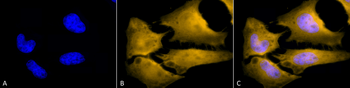
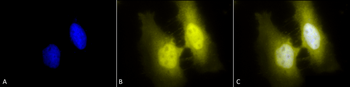
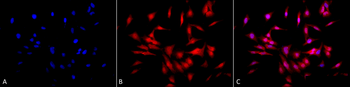
Immunocytochemistry/Immunofluorescence analysis using Rat Anti-HSF1 Monoclonal Antibody, Clone 10H4. Tissue: Heat Shocked cervical cancer cells (HeLa). Species: Human. Fixation: 2% Formaldehyde for 20 min at RT. Primary Antibody: Rat Anti-HSF1 Monoclonal Antibody at 1:100 for 12 hours at 4°C. Secondary Antibody: R-PE Goat Anti-Rat (yellow) at 1:200 for 2 hours at RT. Counterstain: DAPI (blue) nuclear stain at 1:40000 for 2 hours at RT. Localization: Cytoplasm. Localizes to the nucleus upon activation. Magnification: 100x. (A) DAPI (blue) nuclear stain. (B) Anti-HSF1 Antibody. (C) Composite. Heat Shocked at 42°C for 1h.
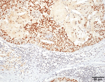
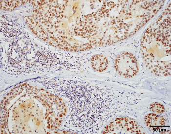
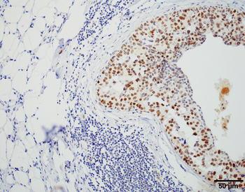
Immunohistochemistry analysis using Rat Anti-HSF1 Monoclonal Antibody, Clone 10H4. Tissue: Breast carcinoma. Species: Human. Fixation: 10% Formalin Solution for 20 hours at RT. Primary Antibody: Rat Anti-HSF1 Monoclonal Antibody at 1:8000 for 40 min. Secondary Antibody: Dako labeled Polymer HRP Anti-rat IgG, DAB Chromogen (brown) (Dako Envision+ System) for 30 min at RT. Counterstain: Mayer's Hematoxylin (purple/blue) nuclear stain for 1 minute at RT. Localization: Nuclear. Magnification: 100X.
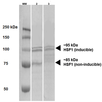
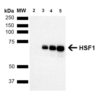
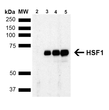
Western Blot analysis of Human A431 and HEK293 cell lysates showing detection of HSF1 protein using Rat Anti-HSF1 Monoclonal Antibody, Clone 10H4. Primary Antibody: Rat Anti-HSF1 Monoclonal Antibody at 1:1000.
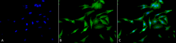
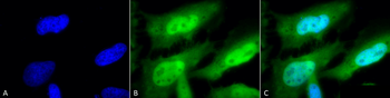
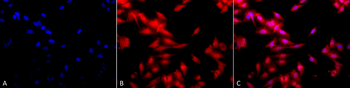
Immunocytochemistry/Immunofluorescence analysis using Rat Anti-HSF1 Monoclonal Antibody, Clone 10H4. Tissue: Heat Shocked cervical cancer cells (HeLa). Species: Human. Fixation: 2% Formaldehyde for 20 min at RT. Primary Antibody: Rat Anti-HSF1 Monoclonal Antibody at 1:100 for 12 hours at 4°C. Secondary Antibody: FITC Goat Anti-Rat (green) at 1:200 for 2 hours at RT. Counterstain: DAPI (blue) nuclear stain at 1:40000 for 2 hours at RT. Localization: Cytoplasm. Localizes to the nucleus upon activation. Magnification: 20x. (A) DAPI (blue) nuclear stain. (B) Anti-HSF1 Antibody. (C) Composite. Heat Shocked at 42°C for 1h.
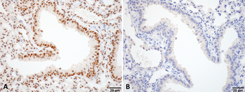
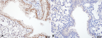
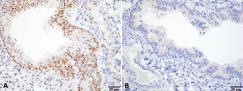
Immunohistochemistry analysis using Rat Anti-HSF1 Monoclonal Antibody, Clone 10H4. Tissue: Lung. Species: Mouse. Fixation: 10% Formalin Solution for 20 hours at RT. Primary Antibody: Rat Anti-HSF1 Monoclonal Antibody at 1:1000 for 40 min. Secondary Antibody: Dako labeled Polymer HRP Anti-rat IgG, DAB Chromogen (brown) (Dako Envision+ System) for 30 min at RT. Counterstain: Mayer's Hematoxylin (purple/blue) nuclear stain for 1 minute at RT. Localization: Nuclear. Magnification: 100X. (A) HSF Wildtype. (B) HSF null.
Quick Database Links
UniProt Details
− No UniProt data available
NCBI Gene Details
− No NCBI Gene data available
NCBI Reference Sequences
−Associated Accession Numbers
Curated reference sequences for the gene transcript and protein product| Protein | NP_032322.1 |
|---|
Documents Download
Datasheet
Product Information
Request a Document
Protocol Information
WB
Western Blot (IB, immunoblot)
IHC
Immunohistochemistry
IF
Immunofluorescence
ICC
Immunocytochemistry
ELISA
Enzyme-linked Immunosorbent Assay (EIA)
IP
Immunoprecipitation
HSF1 Antibody (Biotin) (orb151119)

- 0.0
Based on 0 reviews
Participating in our Biorbyt product reviews program enables you to support fellow scientists by sharing your firsthand experience with our products.
Login to Submit a ReviewAvailable Sizes
Select a size below
Choose Conjugation or Carrier Free Version
Free Secondary Antibody (20 ul)0/0
Please add an antibody product to your cart first.