You have no items in your shopping cart.
Cart summary
Item 1 of 3
Item 1 of 3
Histone H3 antibody
Catalog Number: orb412573
| Catalog Number | orb412573 |
|---|---|
| Category | Antibodies |
| Description | Rabbit polyclonal antibody to Histone H3 |
| Species/Host | Rabbit |
| Clonality | Polyclonal |
| Tested applications | IF, IH, IP, WB |
| Reactivity | Human, Mouse, Rat |
| Immunogen | KLH-conjugated synthetic acetylated peptide corresponding to residues surrounding K27 of human Histone H3 protein. The exact sequence is proprietary. |
| Dilution range | WB: 1:500-2000, IP: 1:50-100 |
| Form/Appearance | Liquid in 0.42% Potassium phosphate, 0.87% Sodium chloride, pH 7.3, 30% glycerol, and 0.01% sodium azide. |
| Conjugation | Unconjugated |
| Target | HIST3H3 |
| Entrez | 8290 |
| UniProt ID | Q16695 |
| Source | Rabbit |
| Storage | Shipped at 4°C. Upon delivery aliquot and store at -20°C for one year. Avoid freeze/thaw cycles. |
| Buffer/Preservatives | Liquid in 0.42% Potassium phosphate, 0.87% Sodium chloride, pH 7.3, 30% glycerol, and 0.01% sodium azide. |
| Alternative names | anti-H3FT antibody, anti-Histone H3.1t antibody, a Read more... |
| Note | For research use only |
| Expiration Date | 12 months from date of receipt. |
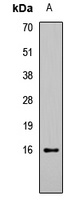
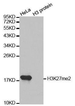
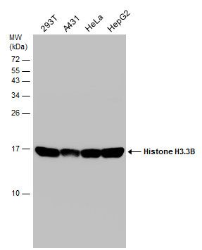
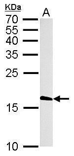
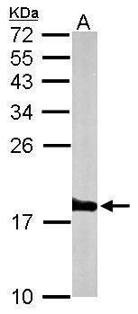
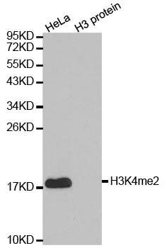
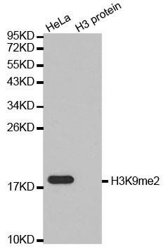
Western blot analysis of Histone H3 (Acetyl-K27) expression in HEK293T (A) whole cell lysates. (Predicted band size: 15 kD; Observed band size: 16 kD)
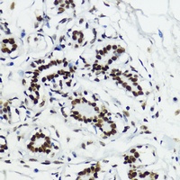
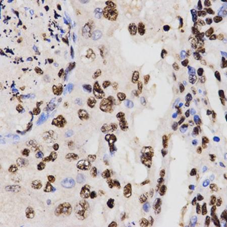
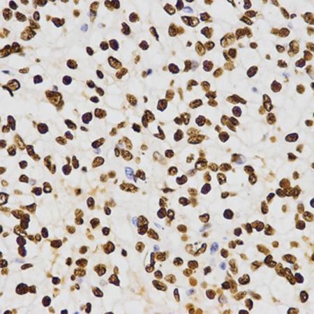
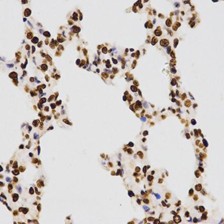
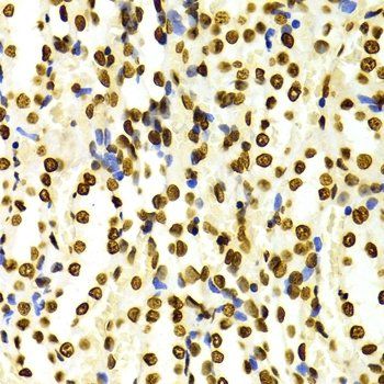
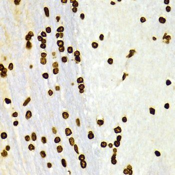
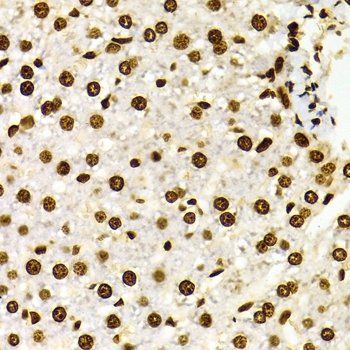
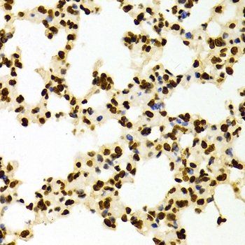
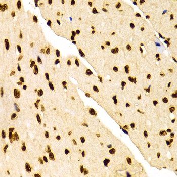
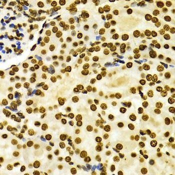
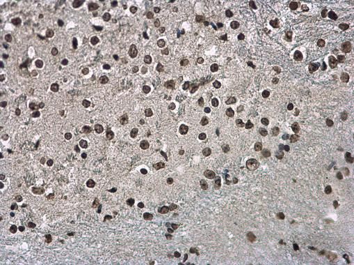
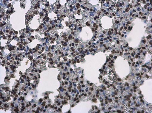
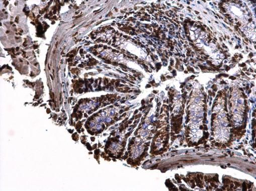
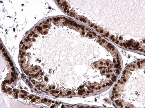
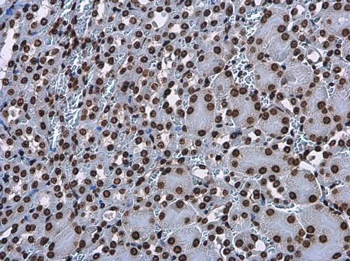
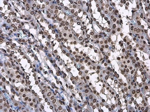
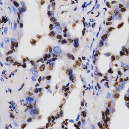
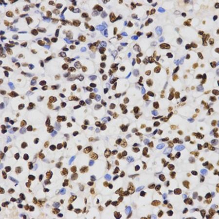
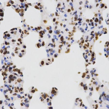
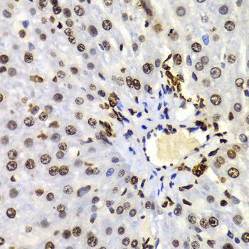
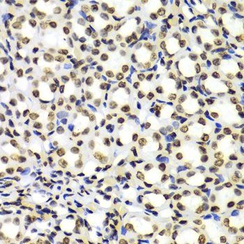
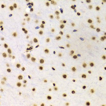
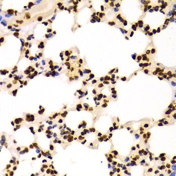
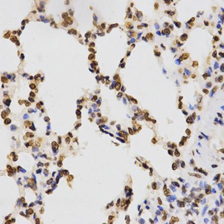
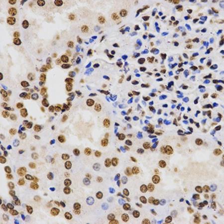
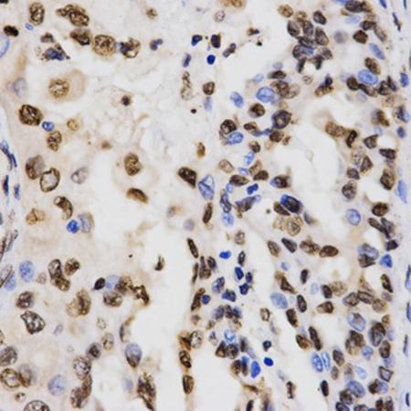
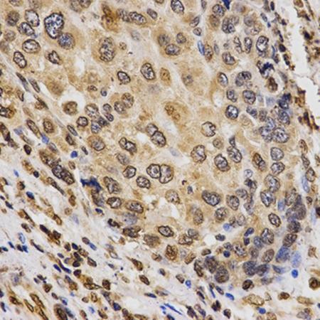
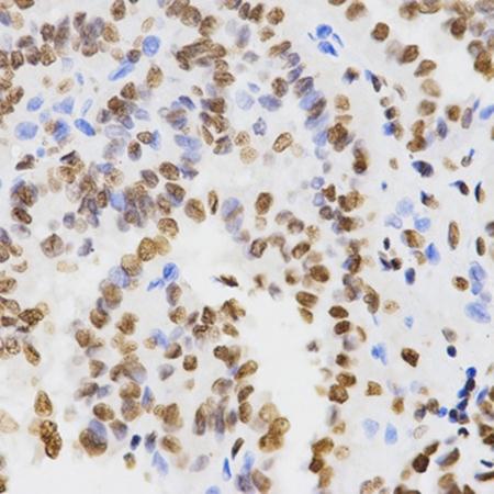
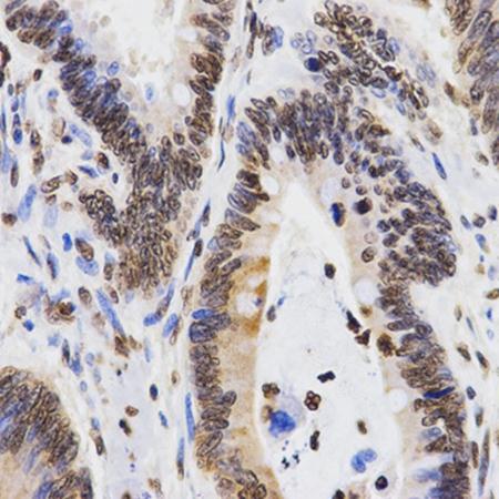
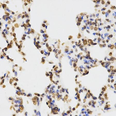
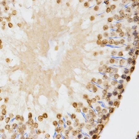
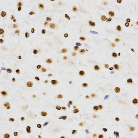
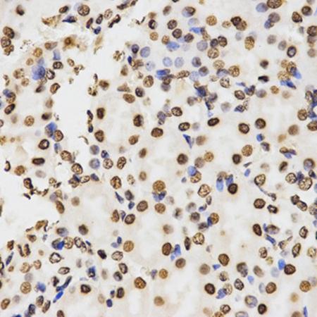
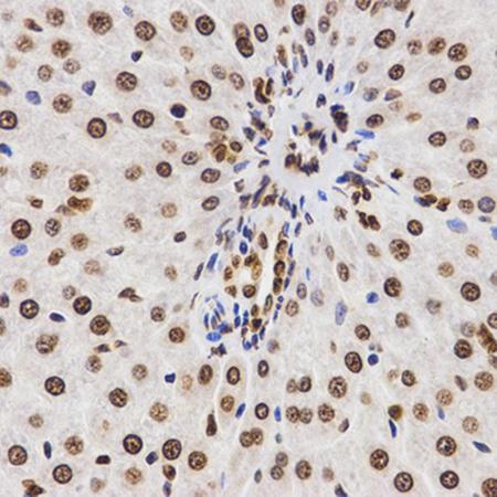
Immunohistochemical analysis of Histone H3 (Acetyl-K27) staining in human breast formalin fixed paraffin embedded tissue section. The section was pre-treated using heat mediated antigen retrieval with sodium citrate buffer (pH 6.0). The section was then incubated with the antibody at room temperature and detected using an HRP conjugated compact polymer system. DAB was used as the chromogen. The section was then counterstained with haematoxylin and mounted with DPX.
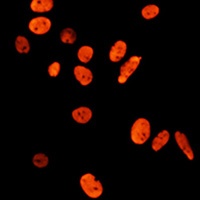
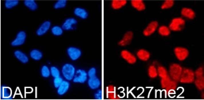
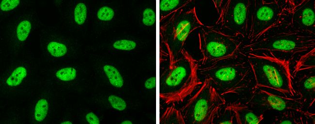
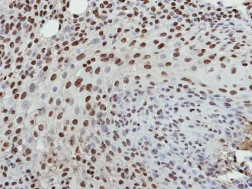
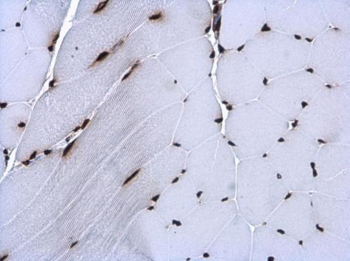
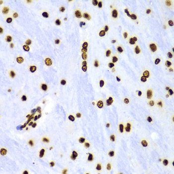
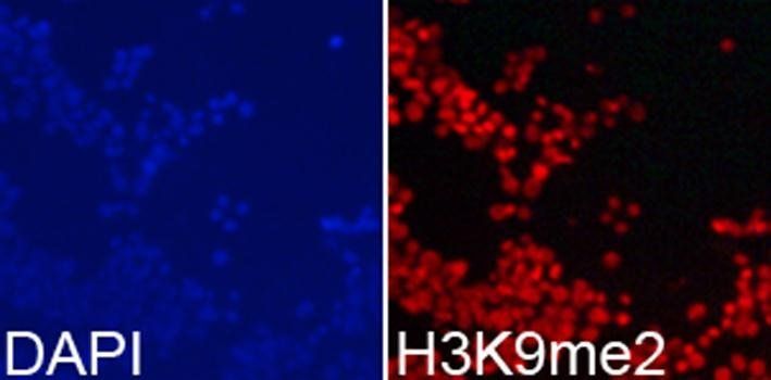
Immunofluorescent analysis of Histone H3 (Acetyl-K27) staining in C6 cells. Formalin-fixed cells were permeabilized with 0.1% Triton X-100 in TBS for 5-10 minutes and blocked with 3% BSA-PBS for 30 minutes at room temperature. Cells were probed with the primary antibody in 3% BSA-PBS and incubated overnight at 4 °C in a humidified chamber. Cells were washed with PBST and incubated with a AF594-conjugated secondary antibody (red) in PBS at room temperature in the dark. DAPI was used to stain the cell nuclei (blue).
- Item 1 of 14
DiMethyl-Histone H3-K27 antibody [orb137139]
ChIP, ICC, IF, IP, WB
All, Human, Mouse, Rat
Unconjugated
50 μl, 100 μl, 200 μl - Item 1 of 13
- Item 1 of 12
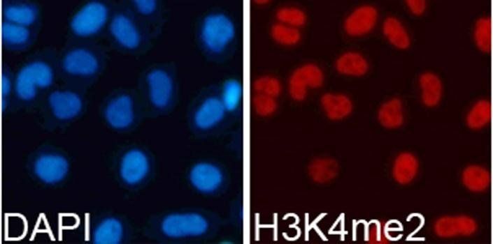
DiMethyl-Histone H3-K4 antibody [orb137148]
ChIP, ICC, IF, IHC, IP, WB
All, Human, Mouse, Rat
Unconjugated
200 μl, 100 μl, 50 μl - Item 1 of 12
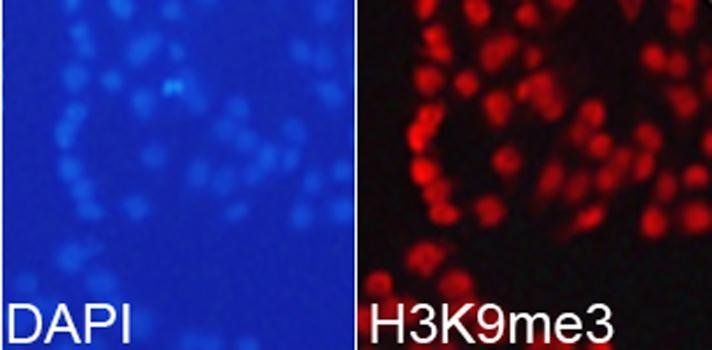
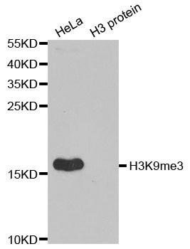
TriMethyl-Histone H3-K9 antibody [orb167161]
ChIP, DOT, ICC, IF, IHC, WB
All, Human, Mouse, Rat
Unconjugated
50 μl, 100 μl, 200 μl - Item 1 of 11
DiMethyl-Histone H3-K9 antibody [orb137153]
ChIP, ICC, IF, IHC, IP, WB
All, Human, Mouse, Rat
Unconjugated
50 μl, 100 μl, 200 μl


Submit a review
Filter by Rating

- 5 stars

- 4 stars
- 3 stars
- 2 stars
- 1 stars