
You have no items in your shopping cart.
Hippo Signaling Pathway
A comprehensive guide to the Hippo signaling cascade, mechanotransduction, YAP/TAZ regulation, and its role in tissue homeostasis and cancer.
The Hippo pathway was first discovered to be involved in controlling tissue growth in Drosophila melanogaster. The major functions of the Hippo pathway restrict tissue growth in adults and modulate cell proliferation, differentiation, and migration in developing organs. Furthermore, dysregulation of the Hippo pathway leads to aberrant cell growth and neoplasia.
The Hippo signalling pathway controls cell proliferation and survival mainly by sensing the physical state of cells within a tissue through several mechanisms. Unlike other classical pathways, Hippo does not have dedicated extracellular ligand–receptor complexes. Instead, the upstream signaling of Hippo involves complex structural mechanotransduction and crosstalk with other signaling pathways.
1. Hippo Signaling Pathway Map
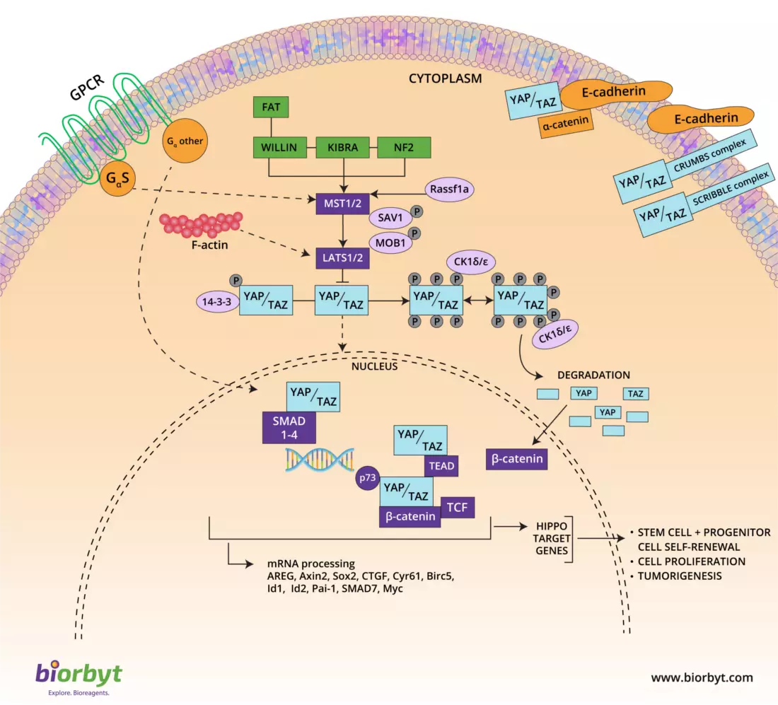
Upstream inputs can generally be divided into two main categories: those controlling the pathway by retaining YAP/TAZ in the cytoplasm through physical interaction, and those that activate the core kinase complex (MST/LATS), leading to YAP/TAZ phosphorylation.
2. The Core Kinase Cascade
The core of Hippo signaling consists of a highly conserved group of serine/threonine kinases and adaptor proteins that tightly regulate the activity of the downstream transcriptional co-activators YAP (Yes-associated protein) and TAZ (transcriptional co-activator with PDZ-binding motif).
A
Kinase Activation (ON State)
When the pathway is active, the kinases MST1/2 (complexed with adaptor protein SAV1) phosphorylate and activate LATS1/2 (complexed with adaptor MOB1). Active LATS1/2 then phosphorylate YAP and TAZ. This phosphorylation creates binding sites for 14-3-3 proteins, which sequester YAP/TAZ in the cytoplasm and target them for proteasomal degradation. This halts cellular proliferation.
B
Kinase Inhibition (OFF State)
If the core kinase complex is inactive, YAP and TAZ remain unphosphorylated. In this state, they avoid degradation and translocate directly into the nucleus. Here, they bind to TEAD family transcription factors, inducing the expression of target genes that promote cell proliferation, survival, and—if dysregulated—cancer development and progression.
3. Upstream Regulation & Mechanotransduction
Because it lacks a dedicated receptor, the Hippo pathway integrates a vast array of intracellular and extracellular structural signals:
- Apical/Basal Polarity & Adhesion: Polarization and adhesion proteins like α-catenin, E-cadherin, CRUMBS, and SCRIBBLE complexes physically sequester YAP/TAZ in the cytoplasm at cell junctions.
- Upstream Core Activators: Proteins including NF2 (Merlin), KIBRA, and WILLIN activate the core kinase complex (MST1/2), keeping cell growth in check.
- GPCR Crosstalk: G-protein-coupled receptors heavily influence Hippo. Signals through Gαs typically lead to the activation of the Hippo cascade (inhibiting YAP), while signals through other Gα subunits (like Gαq/11 or Gα12/13) prefer YAP/TAZ nuclear translocation.
- Mechanotransduction: Mechanical stress regulated by the F-actin cytoskeleton directly impacts YAP/TAZ localization. High mechanical tension (stiff extracellular matrix) typically promotes YAP/TAZ nuclear activity.
Pathological Impact: Loss of upstream contact inhibition (such as loss of E-cadherin) frees YAP/TAZ to enter the nucleus, driving the epithelial-to-mesenchymal transition (EMT), tumor metastasis, and therapeutic resistance.
4. Essential Target Antibodies & Proteins
Biorbyt provides an extensive range of highly specific primary antibodies and recombinant proteins to study both the core kinases and the upstream structural regulators of the Hippo pathway.
| Target Molecule | Description / Pathway Role | Reagent Format |
|---|---|---|
| YAP1 | The primary downstream transcriptional co-activator. Nuclear YAP1 drives proliferation and oncogenesis. | Primary Antibodies, Recombinant Protein |
| TAZ (WWTR1) | A paralog to YAP1, acting synergistically to drive TEAD-mediated gene transcription. | Primary Antibodies |
| MST1 (STK4) & MST2 | The core initiator kinases of the Hippo pathway that phosphorylate LATS1/2. | Primary Antibodies, ELISA Kits |
| LATS1 & LATS2 | The direct kinases responsible for phosphorylating YAP/TAZ, targeting them for degradation. | Primary Antibodies |
| SAV1 (Salvador) | A critical adaptor protein that binds MST1/2 to facilitate full kinase cascade activation. | Primary Antibodies |
| MOB1A / MOB1B | Adaptor proteins that bind to LATS1/2, greatly enhancing their ability to phosphorylate YAP. | Primary Antibodies |
| NF2 (Merlin) | An upstream tumor suppressor that relays contact-inhibition signals to the Hippo core. | Primary Antibodies |
| KIBRA (WWC1) | Forms a complex with NF2 and Ex to activate the MST/LATS cascade at the apical membrane. | Primary Antibodies |
| E-Cadherin | A core component of adherens junctions involved in contact-mediated sequestration of YAP/TAZ. | Primary Antibodies, Recombinant Protein |
| TEAD (1-4) | The family of transcription factors that YAP/TAZ bind to in the nucleus to drive gene expression. | Primary Antibodies |
5. Assays & Small Molecule Modulators
Targeting the Hippo pathway requires precise quantification of YAP/TAZ levels and the ability to modulate their interaction with nuclear transcription factors.
Pathway Inhibition
Verteporfin
A highly characterized small molecule that directly inhibits the interaction between YAP and TEAD, blocking oncogenic transcription.
Quantification
YAP1 ELISA Kits
Quantify total YAP1 expression levels directly from cell lysates or tissue extracts to monitor Hippo pathway suppression.
Quantification
MST1/STK4 ELISA Kits
Highly sensitive assays for the precise measurement of the core Hippo initiator kinase across multiple species.