Cart summary

You have no items in your shopping cart.

Guide to Spatial Biology

What is Spatial Biology?

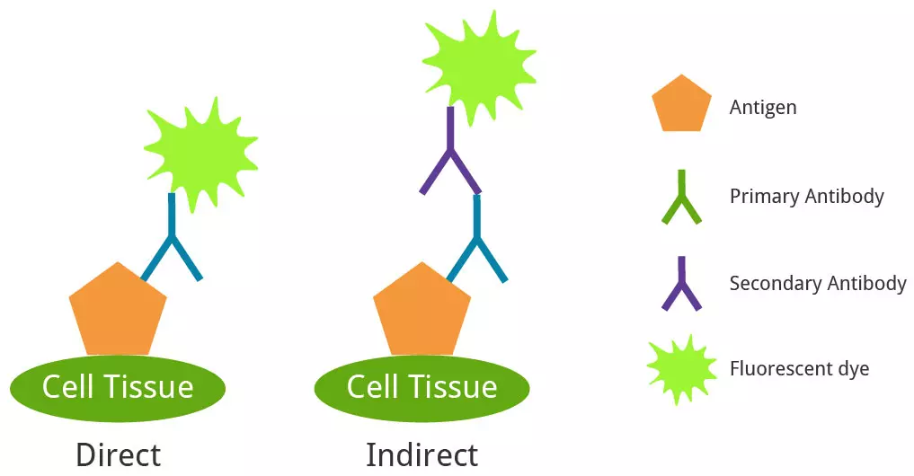
Spatial biology reveals the complex interactions between cells and their surrounding tissue microenvironment—including the extracellular matrix, immune cells, stromal cells, and vasculature. By maintaining spatial context, researchers can gain deeper insights into tissue architecture and disease progression.

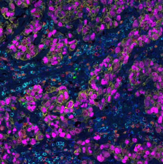
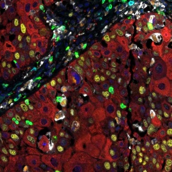
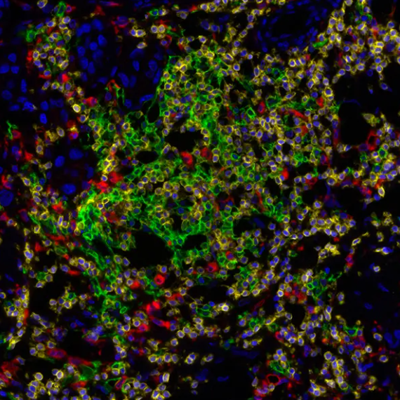
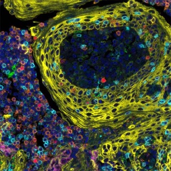
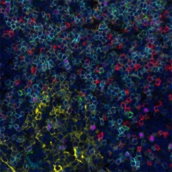
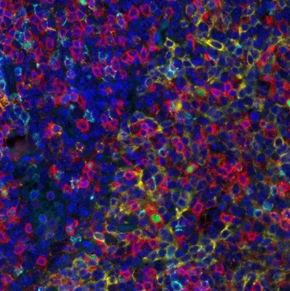
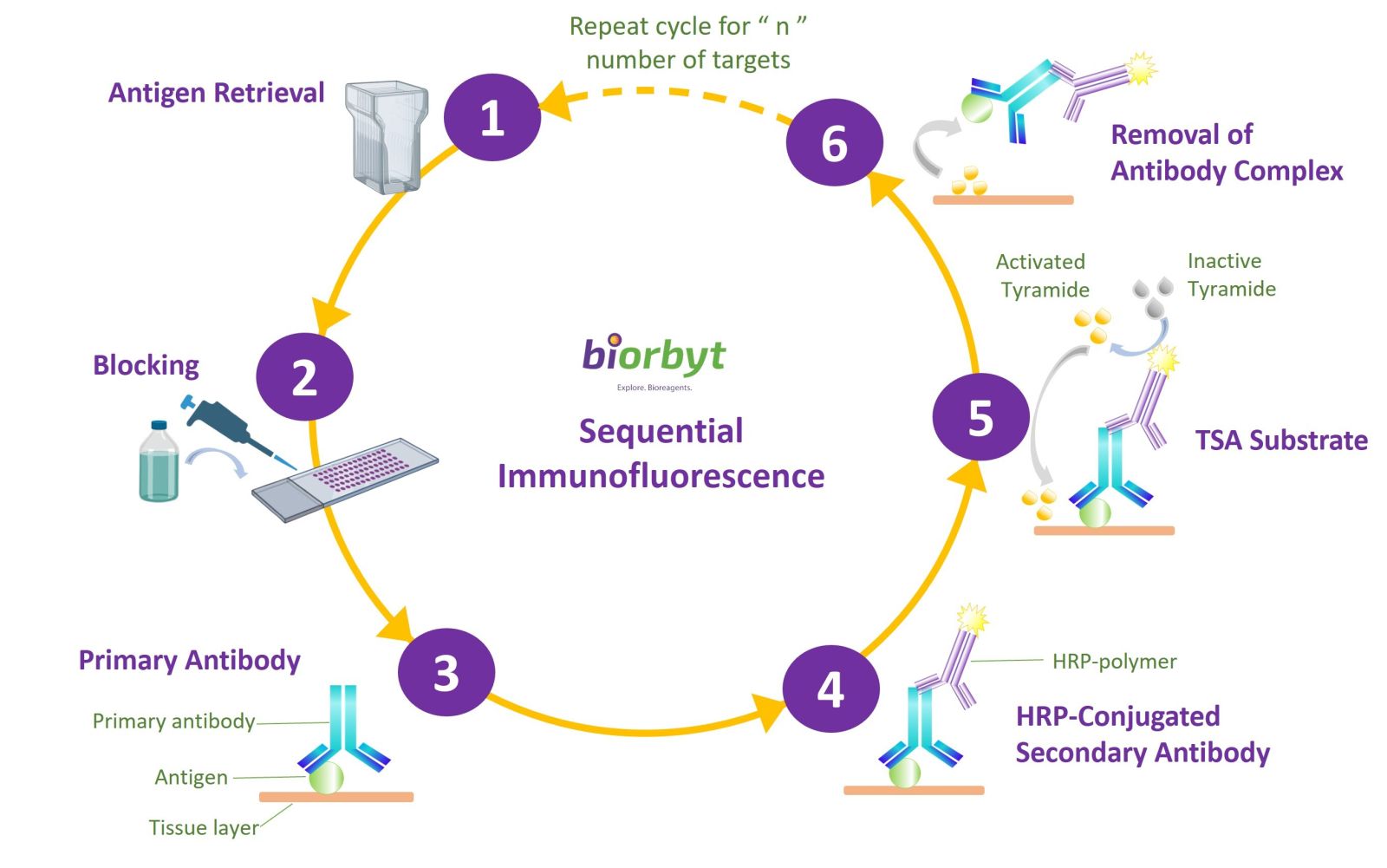
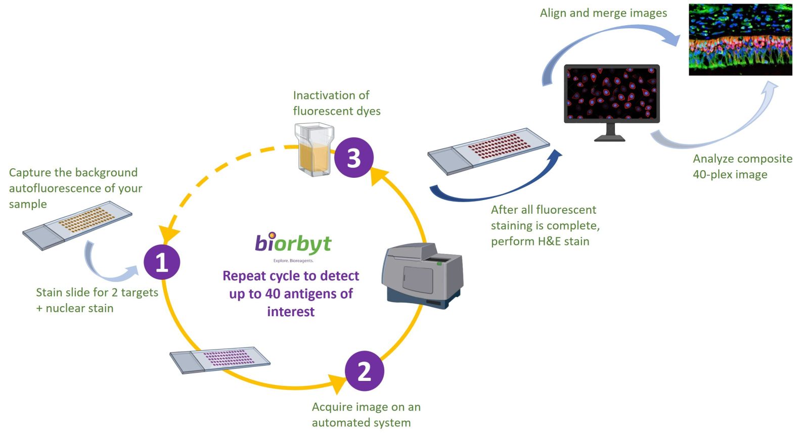
Multiplex Immunofluorescence (mIF)

Multiplex immunofluorescence stands as a vital technique in spatial biology. It allows scientists to detect and study multiple protein markers within a single tissue section—preserving the spatial arrangement and interactions between cells.

Why It Matters?

  • Simultaneous detection of multiple proteins.
  • Detailed mapping of cell types and their interactions.
  • Invaluable for biomarker discovery, therapeutic development, and personalized medicine.

Product ID

Image

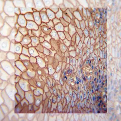
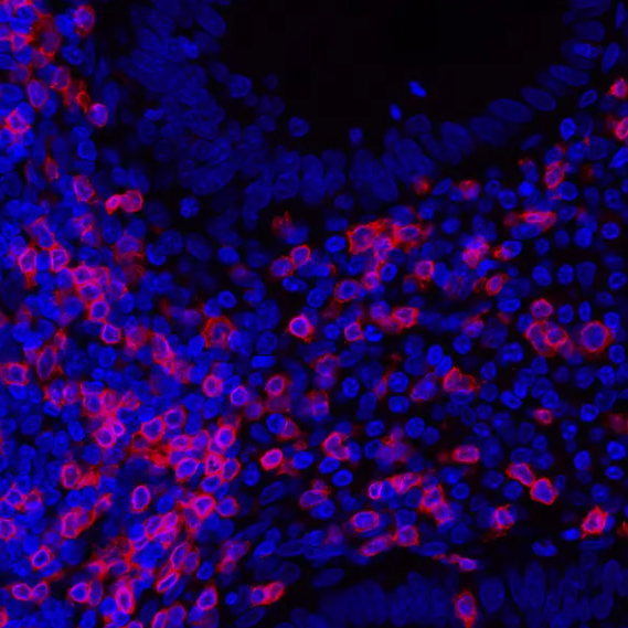
Rabbit anti-CD3E Recombinant Monoclonal Antibody [BL-298-5D12]

Mouse anti-CD20 Monoclonal Antibody, Purified [L26]

Rabbit anti-Ki-67 Recombinant Monoclonal Antibody [BLR021E]

Rabbit anti-PD-L1 Recombinant Monoclonal Antibody [BLR020E]

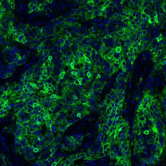
Rabbit anti-Granzyme B Recombinant Monoclonal Antibody [BLR022E]

Tyramide Signal Amplification (TSA)

Tyramide Signal Amplification enhances sensitivity and signal resolution in immunofluorescence staining—perfect for detecting low-abundance targets.

How TSA works

  • HRP-conjugated antibodies and hydrogen peroxide catalyze fluorophore binding to proteins.
  • Primary and secondary antibodies for the first target are applied and deposited.
  • Heat-induced epitope retrieval (HIER) removes them before repeating for the next target.
  • The process is repeated for up to 6 targets (or more), depending on the system.

cycle progress

Does Antibody Order Matter?

The sequence in which antibodies are applied can significantly affect staining quality. Proper ordering ensures optimal antibody performance and minimizes background.

Key reasons include:

  • Heat Exposure: Repeated heating can alter epitope-antibody binding.
  • Tyramide Blocking: Early tyramide deposits can block later antibodies.
  • Tyramide Trapping: Tyramide may trap antibodies, causing unintended signals in later steps.

Cyclic Immunofluorescence (Cyclic IF)

Cyclic IF (also known as CyCIF or MxIF) enables high-plex imaging by cycling through multiple rounds of staining and imaging.

How Cyclic IF Works:

  • Multiple fluorophore-conjugated antibodies are applied in each round.
  • After imaging, dyes are inactivated (via photobleaching or chemical methods).
  • The same fluorophores can then be reused in the next cycle.
  • Images from all cycles are aligned and merged using nuclear staining (DAPI or Hoechst).

Cyclic IF

Advantages:

  • Enables the detection of dozens of targets
  • Maintains tissue and epitope integrity
  • Requires fewer unique fluorophores
  • Allows for detailed spatial analysis

Recommended Products:

Product

Image

Immune Checkpoint PathPlex Panel (CD3E, CD8 alpha, PD-L1)

Cell Proliferation and Metastasis PathPlex Panel (CD3E, Granzyme B, CD8 alpha, Cytokeratin, Ki-67, SOX10)

Activated T Cell PathPlex Panel (CD3E, Cytokeratin, CD8 alpha, CD68, Ki-67, PD-L1)

Immune Localization PathPlex Panel (CD3E, CD68, CD20)

Immunosuppression PathPlex Panel (CD3E, Cytokeratin, CD8 alpha, CD68, PD-L1, FOXP3)

T Cell Status PathPlex Panel (CD3E, Cytokeratin, CD8 alpha, CD4, LAG3, FOXP3)

T Cell Maturation PathPlex Panel (CD8 alpha, Cytokeratin, CD45RO, CD4, FOXP3)


April 2025

Related Pages