You have no items in your shopping cart.
Cart summary
Item 1 of 3
Item 1 of 3
| Catalog Number | orb256550 |
|---|---|
| Category | Antibodies |
| Description | Rabbit polyclonal antibody to GAP43 |
| Species/Host | Rabbit |
| Clonality | Polyclonal |
| Tested applications | IF, IH, WB |
| Reactivity | Human, Mouse, Primate, Rat, Zebrafish |
| Immunogen | KLH-conjugated synthetic peptide encompassing a sequence within the N-term region of human GAP43. The exact sequence is proprietary. |
| Dilution range | WB: 1:500-1000, IHC-P: 1:100-200, IF/ICC: 1:100-500 |
| Form/Appearance | Liquid in 0.42% Potassium phosphate, 0.87% Sodium chloride, pH 7.3, 30% glycerol, and 0.01% sodium azide. |
| Conjugation | Unconjugated |
| Target | GAP43 |
| Entrez | 29423, 14432, 2596 |
| UniProt ID | P07936, P17677, P06837 |
| Source | Rabbit |
| Storage | Shipped at 4°C. Upon delivery aliquot and store at -20°C for one year. Avoid freeze/thaw cycles. |
| Buffer/Preservatives | Liquid in 0.42% Potassium phosphate, 0.87% Sodium chloride, pH 7.3, 30% glycerol, and 0.01% sodium azide. |
| Alternative names | anti Neuromodulin antibody, anti Axonal membrane p Read more... |
| Note | For research use only |
| Expiration Date | 12 months from date of receipt. |
Filter by Applications
Filter by Reactivity
Masoumeh Pourhadi,Hakimeh Zali,Rasoul Ghasemi,Mehrdad Faizi,Faraz Mojab,Mina Soufi Zomorrod Restoring Synaptic Function: How Intranasal Delivery of 3D-Cultured hUSSC Exosomes Improve Learning and Memory Deficits in Alzheimer's Disease Mol Neurobiol, (2023)
Applications
IF
Reactivity
Rat
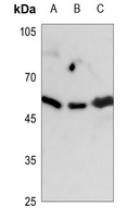
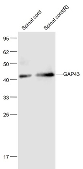
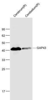
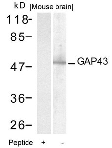
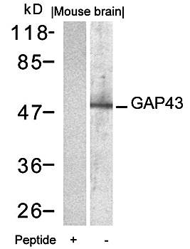
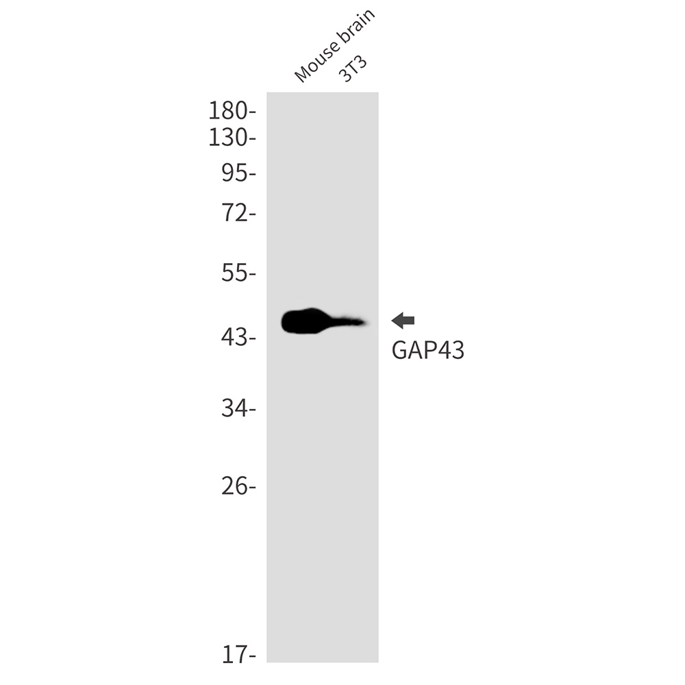
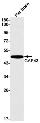
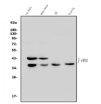
Western blot analysis of GAP43 expression in HEK293T (A), Hela (B), A549 (C) whole cell lysates. (Predicted band size: 24 kD; Observed band size: 48 kD)
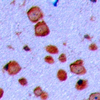
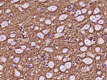
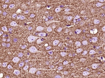
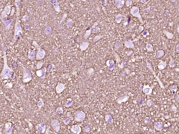
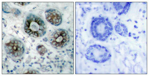
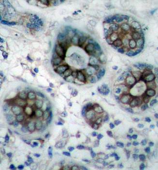
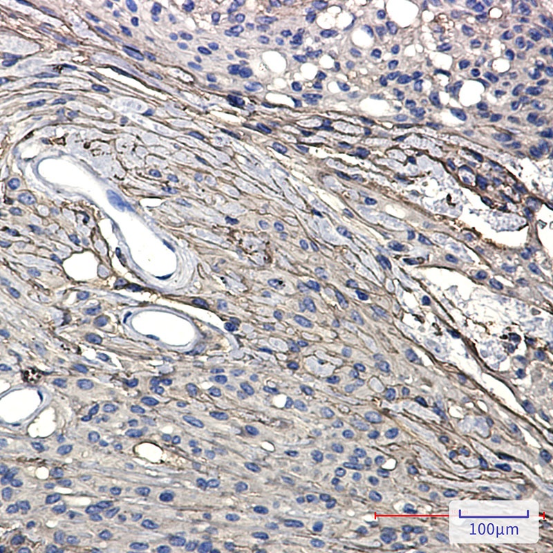
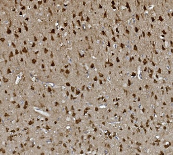
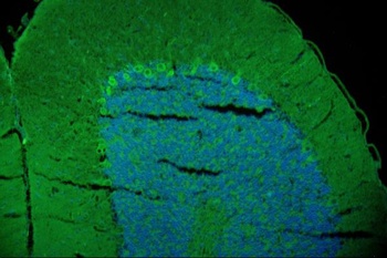
Immunohistochemical analysis of GAP43 staining in human brain formalin fixed paraffin embedded tissue section. The section was pre-treated using heat mediated antigen retrieval with sodium citrate buffer (pH 6.0). The section was then incubated with the antibody at room temperature and detected using an HRP conjugated compact polymer system. DAB was used as the chromogen. The section was then counterstained with haematoxylin and mounted with DPX.
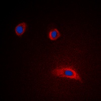
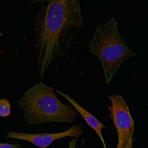
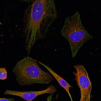
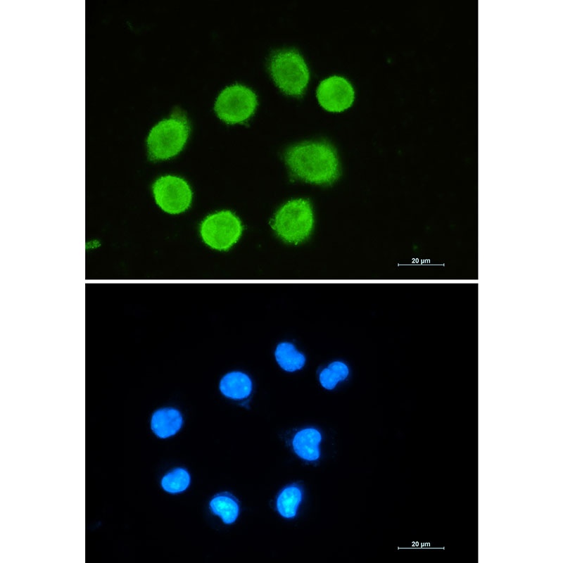
Immunofluorescent analysis of GAP43 staining in HeLa cells. Formalin-fixed cells were permeabilized with 0.1% Triton X-100 in TBS for 5-10 minutes and blocked with 3% BSA-PBS for 30 minutes at room temperature. Cells were probed with the primary antibody in 3% BSA-PBS and incubated overnight at 4 °C in a hidified chamber. Cells were washed with PBST and incubated with a DyLight 594-conjugated secondary antibody (red) in PBS at room temperature in the dark. DAPI was used to stain the cell nuclei (blue).
- Item 1 of 5
- Item 1 of 3
GAP43 (Ab-41) antibody [orb685343]
ELISA, IF, IHC, WB
Human, Mouse, Rat
Rabbit
Polyclonal
Unconjugated
100 μl - Item 1 of 3
GAP43 (Ab-41) Antibody [orb14656]
IF, IHC, WB
Human, Mouse, Rat
Rabbit
Recombinant
Unconjugated
50 μl, 100 μl - Item 1 of 4
GAP43 Antibody [orb1564869]
ICC, IHC-Fr, IHC-P, IP, WB
Human, Mouse, Rat
Rabbit
Monoclonal
Unconjugated
100 μl, 50 μl, 20 μl - Item 1 of 4
GAP43 Antibody [orb654298]
FC, IF, IHC, WB
Human, Mouse, Rat
Rabbit
Polyclonal
Unconjugated
10 μg, 100 μg
Submit a review
Filter by Rating

- 5 stars

- 4 stars
- 3 stars
- 2 stars
- 1 stars