
You have no items in your shopping cart.
Neuroscience Research Tools
Cell Markers in Neuroscience
Identifying and classifying neurons, astrocytes, microglia, and oligodendrocytes utilizing validated target proteins.
Cell markers are specialized functional molecules (typically surface receptors or intracellular structural proteins) that are utilized to accurately identify and systematically classify individual cell populations.
In the dynamic field of neuroscience, critical research into the architecture of the brain and complex nervous systems often inherently requires the precise examination and differentiation of vastly different cell types. This includes mapping intricate networks of neurons, astrocytes, oligodendrocytes, and microglia. Here, we discuss the diverse cell populations of the nervous system and the most common, gold-standard protein markers utilized to reliably identify these cells in vitro and in vivo.
1. Neurons
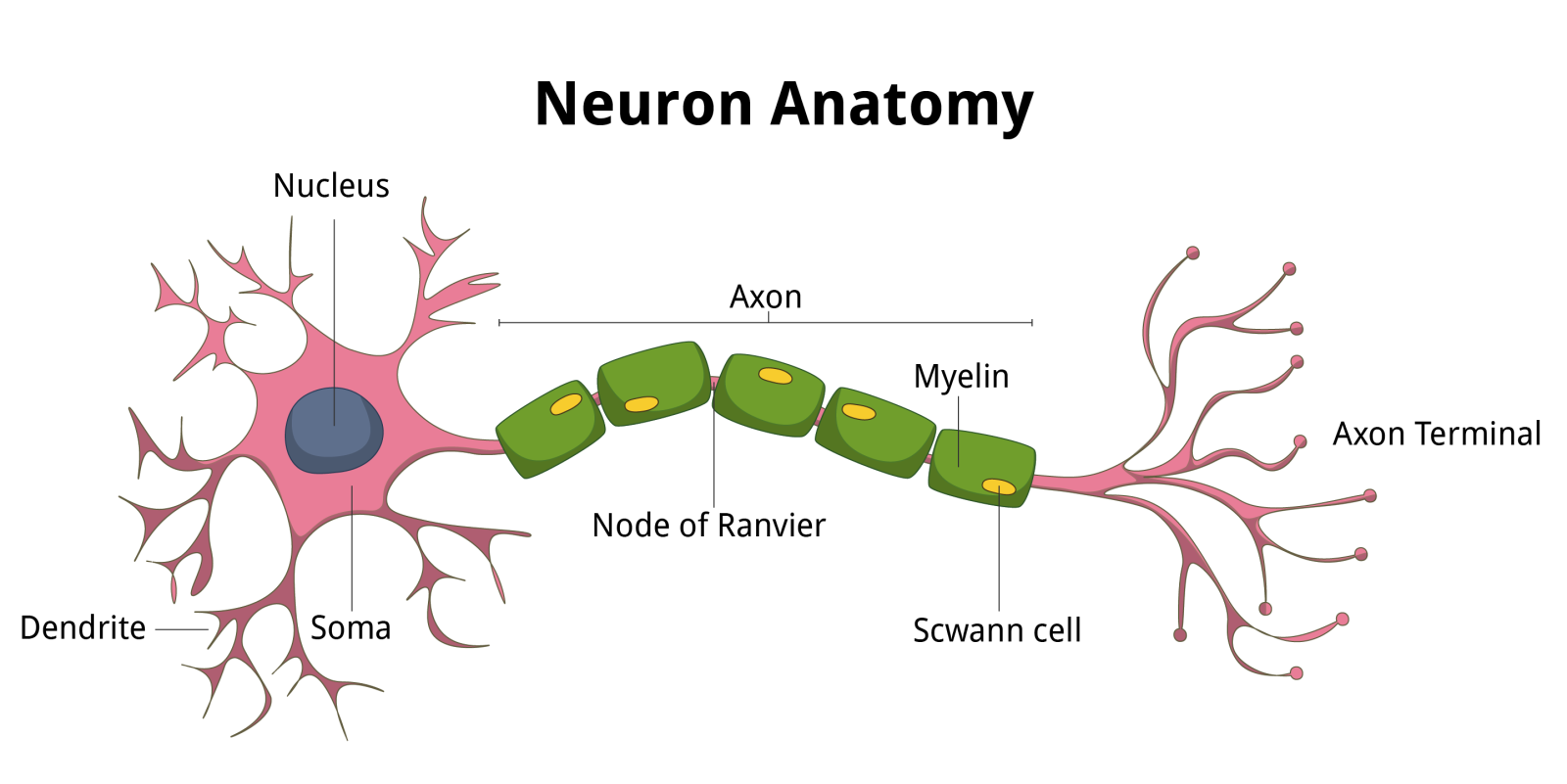
Neurons are the primary, electrically excitable functional cell type of both the central nervous system (CNS) and the peripheral nervous system (PNS). They are exclusively responsible for rapid synaptic communication, functionally allowing us to think, talk, breathe, move, and process complex sensory data.
Neurons communicate dynamically through the targeted transmission and reception of rapid electrical impulses and precise neurochemical signals (neurotransmitters). Structurally, they are distinctly composed of three main architectural parts: the central cell body (soma), the heavily branched dendrites, and the elongated axon. The cell body is responsible for core metabolic functioning, the dendrites physically receive incoming information from upstream neurons, and the axons actively transmit information to downstream cellular targets.
Common Neuronal Markers
Highly specific intracellular protein markers are heavily utilized in neuro-research to confirm distinct neuronal lineages, track cellular maturation, and visualize complex axonal networks. The two primary markers are NeuN and βIII-tubulin.
Neuronal Nuclear Protein (NeuN)
Neuronal nuclear protein, widely known as NeuN, is a specialized regulatory protein absolutely exclusive to mature neurons. This unique protein, found distinctly in both the cellular nucleus and the surrounding cytoplasm, functions as a critical regulatory splicing factor. In neurological research, scientists heavily utilize specific NeuN antibodies to accurately study stem cell differentiation into functional neuronal lineages. In clinical histopathology, scientists are actively utilizing this stable biomarker to assist in complex cancer diagnostics.
→ Explore Biorbyt NeuN Antibodies & Reagents
βIII-tubulin (TUBB3)
βIII-tubulin is a highly specialized microtubule protein structurally encoded by the gene TUBB3. This essential protein plays an immensely important structural role in a wide number of dynamic cellular processes, including early neurogenesis and the aggressive physical regrowth of severed axons and targeted dendrites after traumatic injury. βIII-tubulin is routinely used as the gold-standard biomarker in evaluating neuronal cell differentiation. It is also actively used in the identification of severe neurological disorders, as a genetic mutation in this specific protein has been clinically linked to highly disruptive conditions such as MCD, which can cause severe epilepsy, cerebral palsy, and profound developmental delays.
→ Explore Biorbyt βIII-tubulin Antibodies & Reagents
2. Astrocytes
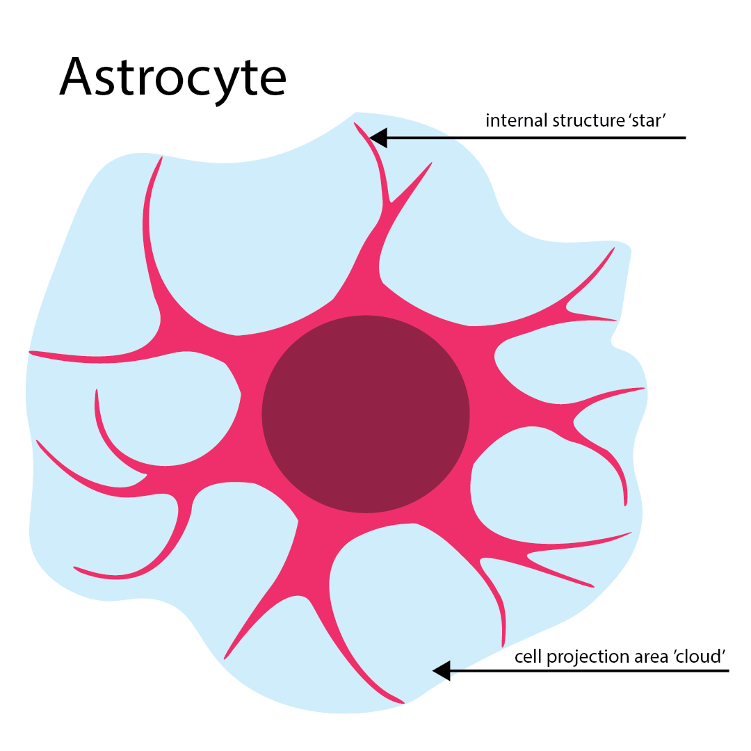
Astrocytes are the most highly prevalent type of supportive glial cell found within the CNS and are densely populated throughout both the brain and the spinal cord. Within the nervous system, astrocytes play a profoundly important role in maintaining chemical homeostasis, the dynamic control of local blood flow, the physical and chemical regulation of synaptic junctions, and ongoing adult neurogenesis.
Pathological loss or functional abnormalities in astrocyte function have been heavily implicated in a wide variety of severe neurodegenerative diseases, actively including Alzheimer's disease, Huntington's disease, and Parkinson’s disease.
Glial Fibrillary Acidic Protein (GFAP)
Glial fibrillary acidic protein (GFAP) is undeniably the most common and reliable astrocyte cell marker. GFAP is an intermediate filament III protein that plays an incredibly important structural role in the intricate cytoskeleton, helping to dynamically maintain cellular strength, shape, and physical support for surrounding neurons. Dramatically increased local levels of GFAP can strongly indicate the active presence of injurious, inflammatory, or neurodegenerative conditions, including traumatic brain injury, aggressive Parkinson’s disease, and advancing Alzheimer’s disease.
→ Explore Biorbyt GFAP Antibodies & Reagents
3. Microglia
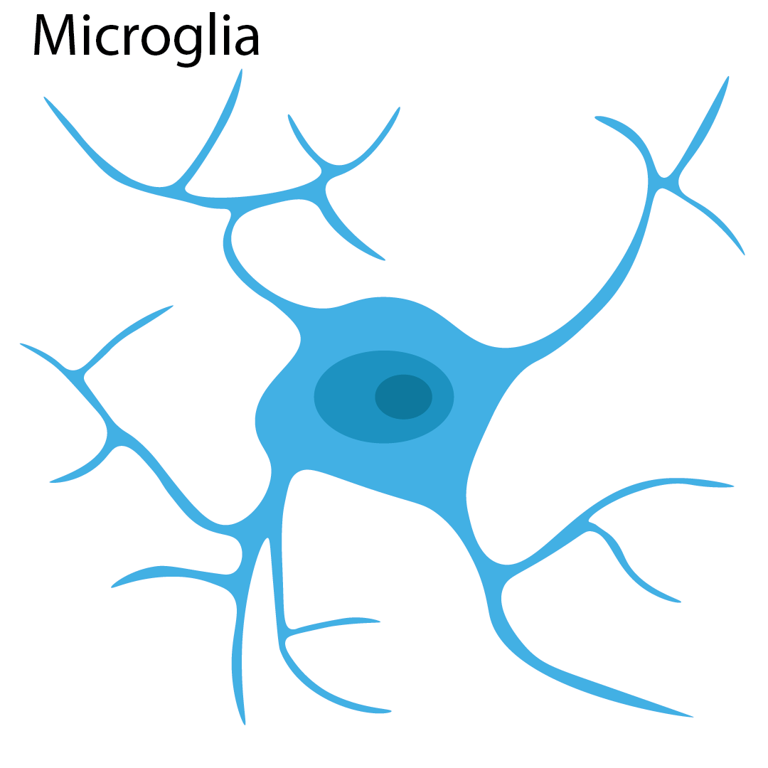
Microglia are a specialized type of active glial cell that definitively serve as the primary resident macrophages and innate immune defense within the mammalian brain. Microglia continuously scavenge the CNS to actively maintain delicate homeostasis by rapidly engulfing and entirely removing harmful substances, such as dead or dying neurons, infectious microbes, and toxic protein aggregates. Microglia also robustly help to regulate complex neuro-inflammation and dynamically prune developing synapses.
Ionized Calcium Binding Adaptor Molecule 1 (Iba1)
Ionized calcium binding adaptor molecule 1 (Iba1) is the absolute gold-standard, common marker for active and resting microglia. Iba1 is a specialized actin-binding protein that heavily plays a functional role in the dynamic restructuring of the cellular cytoskeleton during active phagocytosis and rapid cellular movement toward sites of injury. This highly specific neuromarker can additionally be used clinically to accurately identify dense tumors with a definitive monocytic origin and in the identification of complex histiocytic disorders.
→ Explore Biorbyt Iba1 Antibodies & Reagents
4. Oligodendrocytes
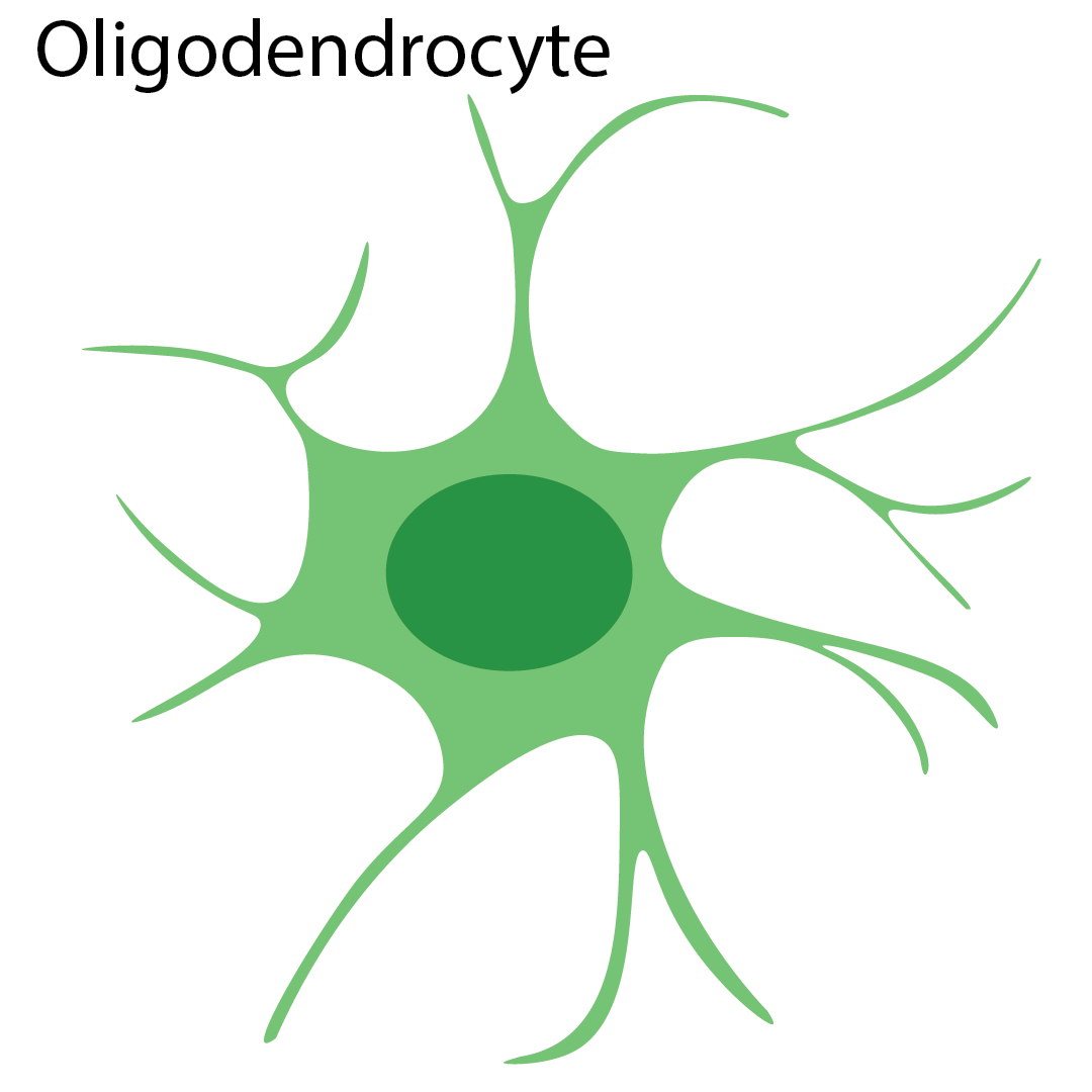
Oligodendrocytes are a highly specialized type of large glial cell that exclusively produces and heavily maintains crucial myelin within the CNS. Myelin dynamically forms the thick, insulating myelin sheaths that tightly surround targeted axons. This critical insulation plays a profoundly important physical role in the strength, structural integrity, and extremely rapid speed of electrical action potential transmission.
Pathological demyelination, or the destructive breakdown of existing myelin sheaths, has been strongly associated with numerous severely debilitating conditions, the most globally well-known being Multiple Sclerosis. There are multiple, highly specific surface biomarkers associated with these unique myelin proteins.
Myelin-Oligodendrocyte Glycoprotein (MOG)
MOG is a specialized structural glycoprotein that is uniquely found on the outermost surface of mature myelin and active oligodendrocyte plasma membranes. MOG is structurally thought to play a vital role in the long-term physical stability of internal oligodendrocyte microtubules and in maintaining the complex, multi-layered architecture of the thick myelin sheaths. Because of its highly exposed surface position, MOG is extremely often used as the primary target in autoimmune system research with respect to severe demyelinating diseases in the central nervous system.
→ Explore Biorbyt MOG Antibodies & Reagents
Myelin-Associated Glycoprotein (MAG)
MAG is a functional transmembrane glycoprotein presenting actively with two distinct structural isoforms: S-MAG (67kDa) and L-MAG (72kDa). MAG acts directly as an adhesion molecule and essentially functions in the highly regulated initiation of myelination, as well as the sustained cellular differentiation and long-term functional maintenance of mature, myelinating oligodendrocytes.
→ Explore Biorbyt MAG Antibodies & Reagents
Myelin Basic Protein (MBP)
MBP is an incredibly abundant structural component that definitively makes up approximately 30% of the total protein found embedded within healthy myelin in the CNS. It is technically classified as an intrinsically disordered protein, meaning it can functionally take on vastly different conformational shapes depending on the local cellular environment. MBP is essentially thought to be a highly multifunctional protein, heavily involved in both tightly compacting myelin structure and complex intracellular signaling cascades.
→ Explore Biorbyt MBP Antibodies & Reagents