You have no items in your shopping cart.
Blotto Immunoanalytical Grade (Non-Fat Dry Milk)
SKU: orb348624
Description
Images & Validation
−Item 1 of 2
| Application Notes |
|---|
Key Properties
−| Purity | This product contains highly purified immunoanalytical grade non-fat dry milk. |
|---|---|
| Conjugation | Unconjugated |
Storage & Handling
−| Storage | Store container at room temperature prior to opening. After reconstitution, use blocking buffers immediately. Dilute solutions may be stored at 4° C for up to four (4) days. Solutions containing BLOTTO may be frozen. |
|---|---|
| Form/Appearance | Lyophilized |
| Buffer/Preservatives | Preservative: None. Stabilizer: None. None |
| Concentration | 1X |
| Expiration Date | 12 months from date of receipt. |
| Hazard Information | Non-Toxic |
| Disclaimer | For research use only |
Alternative Names
−Blotto Immunoanalytical Grade Non-Fat Dry Milk, Blocking reagent for western, antibody dilution buffer
Quality Guarantee
Explore bioreagents carefree to elevate your research. All our products are rigorously tested for performance. If a product does not perform as described on its datasheet, our scientific support team will provide expert troubleshooting, a prompt replacement, or a refund. For full details, please see our Terms & Conditions and Buying Guide. Contact us at [email protected].
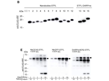
FD-CF Production of On-Demand Affinity Products (B) Anti-FLAG WB showing FD-CF expression of 12 Nanobodies and three DARPins containing ST-FLAG tags (STFL). Specific antigen targets are: (1) CEA5, (2) dengue Virus NS1, (3) GFP, (4) HIV p23-gag, (5) norovirus capsid VP1, (6) rotavirus capsid VP6, (7) C. difficile exotoxin TcdA, (8) P. falciparum VAR2CSA, (9) GLUT1, (10) mCherry, (11) Vimentin, (12) Glycophorin A, (13) HER-2, (14) VEGF-A, and (15) epCAM. (–) indicates a DNA null control reaction. (E) Anti-FLAG WB showing one-pot manufacturing of affinity-output proteins, produced by mixing DNA templates encoding different affinity components (left, anti-CEA5-STFL Nanobody; middle, anti-GFP-STFL Nanobody; right, STFL-anti-HER2 DARPin) with DNA encoding the YFP-SC output component in a single FD-CF reaction. Template ratios are shown below. Membrane was blocked in 4% Blotto (p/n orb348624) +2% cold water fish gelatin.

Screening multiple cell lines reveals that T47D and MCF7 cells efficiently process recombinant MPO into the mature heterotetrameric form. (A) Cell extracts were made from the indicated stable cell lines and the total MPO concentration in each extract quantitated by ELISA. Cell extract containing 2 ng of total MPO was run on Bis-Tris gels in the absence or presence of reducing agent and immunoblotted with a multi-epitope polyclonal MPO antibody to assess the MPO species present in each cell line. Under non-reducing conditions the MPO heterotetramer migrates as three bands between the 100–150 kDa markers. Under reducing conditions, the heterotetramer falls apart and we instead observe the heavy chain (HC) of MPO that migrates just above the 50 kDa marker. Monomeric proMPO migrates just above the 75 kDa marker and shifts slightly upward upon reduction of the intra-molecular disulfide bonds. Untransfected T47D cells (-C) were included as a negative control. (B) The three bands between the 100–150 kDa markers that represent fully processed MPO on non-reducing SDS PAGE were further analyzed to determine if subunit structure was responsible for their distinct gel mobilities. Purified nMPO was run on non-reducing SDS-PAGE and duplicate blots were probed with antibodies specific for either the light chain (L) or heavy chain (H) of MPO. The subunit stoichiometry consistent with the relative immunoreactivity of each band is indicated between the two panels. (C) MPO concentrations in 24 h conditioned media and corresponding whole cell extracts were determined by ELISA and the relative amounts of secreted versus cellular MPO were calculated for each cell line. (D) Whole cell extracts from each cell line were assayed for peroxidase activity and total MPO content and the relative specific activity (Active MPO/Total MPO). (E) Comparison of the sensitivity of native (HL60) and recombinant (T47D) MPO to inactivation by H2O2. Live cells were treated with increasing concentrations of H2O2 in the growth media for 1.5 h at which time further inactivation was stopped by the addition of catalase. Specific peroxidase activity was measured in cell extracts generated from treated cells as in panel D. IC50 values are 2.8 mM (HL60) and 0.96 mM (T47D-MPO). (C, D & E) Assay points are triplicate measurements and plotted as mean ± SE. The data shown is representative of at least three independent experiments. Membranes were blocked with 4% non-fat dry milk and plates were blocked 2% non-fat dry milk (p/n orb348624). (nd) None detected.
Blotto Immunoanalytical Grade (Non-Fat Dry Milk) (orb348624)

- 0.0
Based on 0 reviews
Participating in our Biorbyt product reviews program enables you to support fellow scientists by sharing your firsthand experience with our products.
Login to Submit a ReviewAvailable Sizes
Select a size below