What is Intracellular Cytokine Staining?
Intracellular Cytokine Staining (ICS) is a versatile and powerful technique widely employed in both basic research and preclinical studies to gain comprehensive insights into cellular function and immune responses. This method facilitates the simultaneous assessment of surface markers and intracellular cytokines, providing a multifaceted view of cellular behaviour.
Monensin and Brefeldin A are used in intracellular cytokine staining to trap cytokines inside cells. Monensin disrupts Golgi acidification, causing cytokines to accumulate in the Golgi. Brefeldin A blocks protein transport from the endoplasmic reticulum to the Golgi, leading to cytokine accumulation in the endoplasmic reticulum. Monensin is suited for long-term experiments, while Brefeldin A is ideal for short-term studies.
Why Measure Intracellular Cytokines?
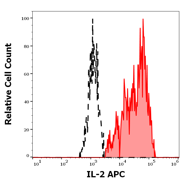
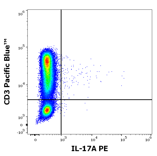
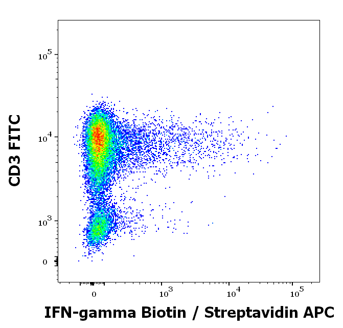
- Surface Marker and Cytokine Measurement: ICS allows researchers to measure cell surface markers, such as CD4 or CD8, which are crucial for identifying different T cell subsets and other immune cell types. At the same time, ICS can quantify intracellular cytokines like IL-2 or IL-17, which are key indicators of cellular activity and immune responses.
- Functional Analysis: By measuring cytokines within cells, researchers can assess the activation status of immune cells, identify cellular signalling pathways, and evaluate the effectiveness of therapeutic interventions.
- Characterising Cell Phenotypes: The dual capability of ICS enables the identification of specific cell populations that produce particular cytokines. For example, it can reveal which subsets of T cells are responsible for secreting cytokines associated with various immune responses or diseases.
How Does Flow Cytometry Fit In?
- Flow Cytometry is a powerful analytical technology that enables the simultaneous measurement of multiple characteristics of cells as they move in a fluid stream through a laser beam.
- It allows for the concurrent assessment of multiple parameters, including cytokine production, cell size, granularity, and surface marker expression.
- Single-Cell Resolution: This technology provides data at the single-cell level, allowing for a granular view of cellular responses and an understanding of heterogeneous cell populations.
- It facilitates precise quantification of cytokine levels, providing accurate and reproducible data through the use of fluorochrome-conjugated antibodies that specifically bind to target cytokines.
Enhance your Intracellular Cytokine Detection with Biorbyt
Here are some of the most commonly studied cytokines from our catalog:
Data from Biorbyt Products
Select the Right Buffer for Flow Cytometry
If you have any questions, please contact our Technical Team at [email protected].